Download
1 / 91
940 likes | 1.08k Views
METABOLISM OF LIPIDS: DIGESTION OF LIPIDS. TRANSPORT FORMS OF LIPIDS. PHYSIOLOGICAL ROLE OF LIPIDS. Energetic role (fuel molecules) Components of membranes (structural role) Precursors for many hormones (steroids) Signal molecules (prostaglandins)

E N D
METABOLISM OF LIPIDS:DIGESTION OF LIPIDS. TRANSPORT FORMS OF LIPIDS
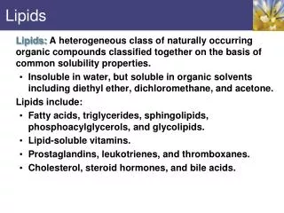
PHYSIOLOGICAL ROLE OF LIPIDS • Energetic role(fuel molecules) • Components of membranes(structural role) • Precursors for many hormones(steroids) • Signal molecules(prostaglandins) • Protective role(lipids surround important organs) • Enzyme cofactors(vitamin K) • Electron carriers(ubiquinone) • Insulation against temperature extremes
TRIACYLGLYCEROLS ARE HIGHLY CONCENTRATED ENERGY STORES • Triacylglycerols (TGs) and glycogen -two major forms of stored energy TGs which are more efficient energy stores because:(1) They are stored in an anhydrous form (2) Their fatty acids are more reduced than monosaccharides.
1 g of triacylglycerols stores more than six times as much energy as a 1 g of glycogen • Glycogen reserves are depleted in 12 to 24 hours after eating, triacylglycerols within several weeks. • Fat breakdown about 50 % of energy in liver, kidney and skeletal muscles up to 95 % of energy cardiac muscle • Fats are the major source of energy for: fasting animal organism in diabetes
Fatty acids and glycerol - substances that are directly used as a fuel by mammalian organisms. • Fatty acids (FA) and glycerol for metabolic fuels are obtained from triacylglycerols: (1) In the diet (2) Stored in adipocytes (fat storage cells) • Free fatty acids occur only in trace amounts in cells • For supplying of fatty acids as a fuel for organism, the triacylglycerols have to be digested
DIGESTION OF DIETARY LIPIDS Lipids in diet:triacylglycerols phospholipids cholesterol Digestion – in small intestine. Enzyme – pancreaticlipase. Lipase catalyzes hydrolysis at the C1 and C3 positions of TGs producing free fatty acids and 2-monoacylglycerol.
Colipase – protein which is present in the intestine and helps bind the water-soluble lipase to the lipid substrates. Colipase also activates lipase. Bile salts (salts of bile acids) are required for lipids digestion. Bile salts are synthesized in the liver from cholesterol. Taurocholate and glycocholate - the most abundant bile salts. Amphipathic: hydrophilic (blue) and hydrophobic (black)
TGs are water insoluble and lipase is water soluble. Digestion of TGs takes place at lipid-water interfaces. Rate of digestion depends on the surface area of the interface. Bile salts are amphipathic, they act as detergent emulsifying the lipid drops and increasing the surface area of the interface.
Bilesalts also activates the lipase. Inadequate production of bile salts results in steatorrhea.
Dietary phospholipids are degraded by phospholipases Phospholipases are synthesized in the pancreas. Major phospholipase is phospholipase A2(catalyses the hydrolysis of ester bond at C2 of glycerophospholipids and lysophosphoglycerides are formed). Lysophospho-glycerides are absorbed and in the intestinal cells are reesterified back to glycero-phospholipids.
Lysophosphoglycerides can act as detergent and therefore in high concentration can disrupt cellular membranes. Lysophosphoglyceride is normally present in cells in low concentration. Snake venom contain phospholipase A2 and causes the lysis of erythrocytes membranes.
Dietary cholesterol • Most dietary cholesterol is unesterified • Cholesteryl esters are hydrolyzed in the intestine by an intestinal esterase • Free cholesterol is solublized by bile-salt micelles for absorption • After absorption in the intestinal cells cholesterol react with acyl-CoA to form cholesteryl ester.
ABSORPTION OF DIETARY LIPIDS Lipid absorption – passive diffusion process. 2-monoacylglycerols, fatty acids, lysophosphoglycerides, free cholesterol form micelles with bile salts.
Micelles migrate to the microvilli and lipids diffuse into the cells. Bile acids are actively absorbed and transferred to the liver via portal vein. Bile salts can circulate through intestine and liver several time per day.
In the intestinal cells the fatty acids are converted to fatty acyl CoA molecules. Three of these molecules can combine with glycerol, or two with monoacylglycerol, to form a triacylglycerols. 1. 2. 1-st reaction is catalyzed by monoacylglycerol acyltransferase 2-nd reaction is catalyzed by diacylglycerol acyltransferase
TRANSPORT FORMS OF LIPIDS • TGs, cholesterol and cholesterol esters are insoluble in water and cannot be transported in blood or lymph as free molecules • These lipids assemble with phospholipids and apoproteins (apolipoproteins) to form spherical particles called lipoprotein Structure: Hydrophobic core: -TGs, -cholesteryl estersHydrophilic surfaces: -cholesterol, -phospholipids, -apolipoproteins
The main classes of lipoproteins • Chylomicrons. • Very low density lipoproteins (VLDL). • Intermediate density lipoproteins (IDL). • Low density lipoproteins (LDL). • High density lipoproteins (HDL).
Lymphatic vessel exocytosis Chylomicrons • are the largest lipoproteins (180 to 500 nm in diameter) • are synthesized in the ERof intestinal cells • contain 85 % of TGs (it is the main transport form of dietary TGs). • apoprotein B-48(apo B-48)is the main protein component • deliver TGs from the intestine (via lymph and blood) to tissues (muscle for energy, adipose for storage). • bind to membrane-bound lipoproteinlipase (at adipose tissue and muscle), where the triacylglycerols are again degraded into free fatty acids and monoacylglycerol for transport into the tissue • are present in blood only after feeding
triacylglycerol cholesteryl esters Apo B Apo E phospholipids cholesterol VLDL • are formed in the liver • contain 50 % of TGs and 22 % of cholesterol • two lipoproteins — apo B-100 and apo E • the main transport form of TGs synthesized in the organism (liver) • deliver the TGs from liver to peripheral tissue (muscle for energy, adipose for storage) • bind to membrane-bound lipoproteinlipases (triacylglycerols are again degraded into free fatty acids and monoacylglycerol)
Lipoproteinlipase – enzyme which is located within capillaries of muscles and adipose tissue Function:hydrolyses of TGs of chylomicrons and VLDL. Formed free fatty acids and glycerol pass into the cells Chylomicrons and VLDL which gave up TGs are called remnants of chylomicrons and remnants of VLDL Remnants are rich in cholesterol esters Remnants of chylomicrons are captured by liver Remnants of VLDL are also called intermediate density lipoproteins (IDL) Fate of the IDL: - some are taken by the liver- others are degraded to thelow density lipoproteins (LDL)(by the removal of more triacylglycerol)
LDL LDL are formed in the blood from IDL and in liver from IDL (enzyme – liver lipase) LDL are enriched in cholesterol and cholesteryl esters (contain about 50 % of cholesterol) Protein component - apo B-100 LDL is the major carrier of cholesterol(transport cholesterol to peripheral tissue)
Cells of all organs have LDL receptors Receptors for LDL are localized in specialized regions called coated pits, which contain a specialized protein called clathrin Apo B-100 on the surface of an LDL binds to the receptor Receptor-LDL complex enters the cell by endocytosis. Endocytic vesicle is formed
Vesicle fuse with lysosomes Lysosomal lipases and proteases degrade LDL LDL receptor itself returns to the plasma membrane Apo B-100 is hydrolyzed to amino acids Cholesteryl esters are hydrolyzed to free cholesterol and fatty acids Released free cholesterol: - is incorporated into the membranes or - is reesterified for storage inside the cell by the enzyme acyl CoA:cholesterol acyltransferase (ACAT) Feedback regulation:abundance of intracellular cholesterol suppresses the synthesis of LDL receptors and so the uptake of additional cholesterol from plasma LDL is blocked
Familial hypercholesterolemia • congenital disease when LDL receptor are not synthesized (mutation at a single autosomal locus) • the concentration of cholesterol in blood markedly increases • severe atherosclerosis is developed (deposition of cholesterol in arteries) • nodules of cholesterol called xanthomas are prominent in skin and tendons • most homozygotes die of coronary artery disease in childhood • the disease in heterozygotes (1 in 500 people) has a milder and more variable clinical course atherosclerosis xanthomas
HDL • are formed in the liver and partially in small intestine • contain the great amount of proteins (about 40 %) • pick up the cholesterol from peripheral tissue, chylomicrons and VLDL • enzyme acyltransferase in HDL esterifies cholesterols, convert it to cholesterol esters and transport to the liver
High serum levels of cholesterol cause disease and death by contributing to development of atherosclerosis Cholesterol which is present in the form of the LDL is so-called "bad cholesterol." Cholesterol in the form of HDL is referred to as "good cholesterol” HDLfunctions as a shuttle that moves cholesterol throughout the body
LDL/HDL Ratio The ratio of cholesterol in the form of LDL to that in the form of HDL can be used to evaluate susceptibility to the development of atherosclerosis For a healthy person, the LDL/HDL ratio is 3.5
LIPID METABOLISM: MOBILIZATION OF TRIACYLGLYCEROLS; OXIDATION OF GLYCEROL
adipocyte Storage and Mobilization of Fatty Acids (FA) • TGs are delivered to adipose tissue in the form of chylomicrones and VLDL, hydrolyzed by lipoprotein lipaseinto fatty acids and glycerol, which are taken up by adipocytes. • Then fatty acids are reesterified to TGs. • TGs are stored in adipocytes. • To supply energy demands fatty acids and glycerol are released – mobilisation of TGs.
At low carbohydrate and insulin concentrations (during fasting), TG hydrolysis is stimulated by epinephrine, norepinephrine, glucagon,andadrenocorticotropic hormone. TG hydro-lysis is inhibited by insulin in fed state
Lipolysis - hydrolysis of triacylglycerols by lipases. • A hormone-sensitive lipase converts TGs to free fatty acids and monoacylglycerol • Monoacylglycerol is hydrolyzed to fatty acid and glycerol or by a hormone-sensitive lipase or by more specific and more active monoacylglycerol lipase
Transport of Fatty Acids and Glycerol • Fatty acids and glycerol diffuse through the adipocyte membrane and enter bloodstream. • Glycerol is transported via the blood in free state and oxidized or converted to glucose in liver. • Fatty acids are traveled bound to albumin. • In heart, skeletal muscles and liver they are oxidized with energy release.
Oxidation of Glycerol Glycerol is absorbed by the liver. Steps: phosphorylation, oxidation and isomerisation. Glyceraldehyde 3-phosphate is an intermediate in: • glycolytic pathway • gluconeogenic pathways
ATP Generation from Glycerol Oxidation glycerol – glycerol 3-phosphate - 1 ATP glycerol 3-phosphate - dihydroxyaceton phosphate 2.5ATP (1 NADH) glyceraldehyde 3-phosphate – pyruvate 4,5 ATP (1NADH + 2 ATP) pyruvate – acetyl CoA 2.5 ATP (1 NADH) acetyl CoA in Krebs cycle 10 ATP (3NADH + 1 FADH2 + 1GTP) Total 19,5-1 = 18,5 ATP
LIPID METABOLISM: FATTY ACID OXIDATION
Stages of fatty acid oxidation (1) Activation of fatty acids takes place on the outer mitochondrial membrane (2) Transport into the mitochondria (3) Degradation to two-carbon fragments (as acetyl CoA) in the mitochondrial matrix(b-oxidation pathway)
(1) Activation of Fatty Acids • Fatty acids are converted to CoA thioesters by acyl-CoA synthetase (ATP dependent) • The PPi released is hydrolyzed by a pyrophosphatase to 2 Pi • Two phosphoanhydride bonds (two ATP equivalents)are consumed to activate one fatty acid to a thioester
(2) Transport of Fatty Acyl CoA into Mitochondria • The carnitine shuttle system. • Fatty acyl CoA is first converted to acylcarnitine (enzymecarnitine acyltransferase I(bound to the outer mitochondrial membrane). • Acylcarnitine enters the mitochondria by a translocase. • The acyl group is transferred back to CoA (enzyme - carnitine acyltransferase II).
Carnitine shuttle system • Path of acyl group in red
(3) The Reactions of b oxidation • The b-oxidation pathway (b-carbon atom (C3) is oxidized) degrades fatty acids two carbons at a time
1.Oxidation of acyl CoA by an acyl CoA dehydrogenase to give an enoyl CoA Coenzyme - FAD
2. Hydration of the double bond between C-2 and C-3 by enoyl CoA hydratase with the 3-hydroxyacyl CoA (b-hydroxyacyl CoA) formation
3. Oxidation of 3-hydroxyacyl CoA to 3-ketoacyl CoA by 3-hydroxyacyl CoA dehydrogenase Coenzyme – NAD+
4. Cleavage of 3-ketoacyl CoA by the thiol group of a second molecule of CoA with the formation of acetyl CoA and an acyl CoA shortened by two carbon atoms. Enzyme - b-ketothiolase.
The shortened acyl CoA then undergoes another cycle of oxidation The number of cycles: n/2-1, where n – the number of carbon atoms
b-Oxidation of saturated fatty acids Fatty acyl CoA
One round of b oxidation: 4 enzyme steps produce acetyl CoA from fatty acyl CoA • Each round generates one molecule each of: FADH2NADHAcetyl CoA Fatty acyl CoA (2 carbons shorter each round) Fates of the products of b-oxidation: - NADH and FADH2 - are used in ETC - acetyl CoA - enters the citric acid cycle - acyl CoA – undergoes the next cycle of oxidation