Download
1 / 1
10 likes | 97 Views
A. Reagent only. Reagent only. B. Sialic Acid Standards. Sialic Acid Standards. 2 days old. C. .05. e. e. b. b. d. d. R. 0.20. 0.20. a. a. R. Fresh. 0.0. 0.15. 0.15. A2 glycan Standard. DGP. R. c. c. 0.2. 0.10. 0.10. f. f. 0.1. 0.05. 0.05. R. 32-kDa Enamelin.

E N D
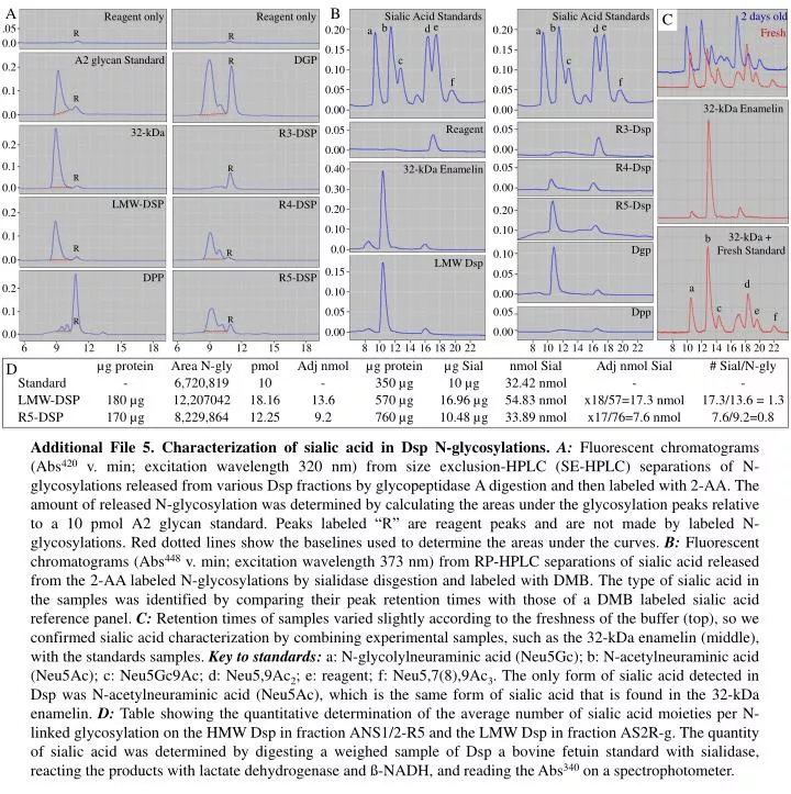
A Reagent only Reagent only B Sialic Acid Standards Sialic Acid Standards 2 days old C .05 e e b b d d R 0.20 0.20 a a R Fresh 0.0 0.15 0.15 A2 glycan Standard DGP R c c 0.2 0.10 0.10 f f 0.1 0.05 0.05 R 32-kDa Enamelin 0.0 0.00 0.00 32-kDa R3-DSP Reagent R3-Dsp 0.05 0.05 0.2 0.00 0.00 0.1 R R4-Dsp 0.05 0.40 32-kDa Enamelin R 0.0 0.00 0.30 LMW-DSP R4-DSP R5-Dsp 0.2 0.20 0.20 0.10 0.1 0.10 32-kDa + Fresh Standard b R R 0.0 Dgp 0.0 0.10 LMW Dsp R5-DSP DPP 0.15 0.05 0.2 d a 0.10 0.00 0.1 c e 0.05 Dpp 0.05 R R f 0.0 0.00 0.00 6 6 9 9 12 12 15 15 18 18 8 10 12 14 16 18 20 22 8 10 12 14 16 18 20 22 8 10 12 14 16 18 20 22 µg protein Area N-gly pmol Adj nmol µg protein µg Sial nmol Sial Adj nmol Sial # Sial/N-gly D Standard - 6,720,819 10 - 350 µg 10 µg 32.42 nmol - - LMW-DSP 180 µg 12,207042 18.16 13.6 570 µg 16.96 µg 54.83 nmol x18/57=17.3 nmol 17.3/13.6 = 1.3 R5-DSP 170 µg 8,229,864 12.25 9.2 760 µg 10.48 µg 33.89 nmol x17/76=7.6 nmol 7.6/9.2=0.8 Additional File 5. Characterization of sialic acid in Dsp N-glycosylations. A:Fluorescent chromatograms (Abs420 v. min; excitation wavelength 320 nm) from size exclusion-HPLC (SE-HPLC) separations of N-glycosylations released from various Dsp fractions by glycopeptidase A digestion and then labeled with 2-AA. The amount of released N-glycosylation was determined by calculating the areas under the glycosylation peaks relative to a 10 pmol A2 glycan standard. Peaks labeled “R” are reagent peaks and are not made by labeled N-glycosylations. Red dotted lines show the baselines used to determine the areas under the curves. B: Fluorescent chromatograms (Abs448 v. min; excitation wavelength 373 nm) from RP-HPLC separations of sialic acid released from the 2-AA labeled N-glycosylations by sialidase disgestion and labeled with DMB. The type of sialic acid in the samples was identified by comparing their peak retention times with those of a DMB labeled sialic acid reference panel. C: Retention times of samples varied slightly according to the freshness of the buffer (top), so we confirmed sialic acid characterization by combining experimental samples, such as the 32-kDa enamelin (middle), with the standards samples. Key to standards: a: N-glycolylneuraminic acid (Neu5Gc); b: N-acetylneuraminic acid (Neu5Ac); c: Neu5Gc9Ac; d: Neu5,9Ac2; e: reagent; f: Neu5,7(8),9Ac3. The only form of sialic acid detected in Dsp was N-acetylneuraminic acid (Neu5Ac), which is the same form of sialic acid that is found in the 32-kDa enamelin. D: Table showing the quantitative determination of the average number of sialic acid moieties per N-linked glycosylation on the HMW Dsp in fraction ANS1/2-R5 and the LMW Dsp in fraction AS2R-g. The quantity of sialic acid was determined by digesting a weighed sample of Dsp a bovine fetuin standard with sialidase, reacting the products with lactate dehydrogenase and ß-NADH, and reading the Abs340 on a spectrophotometer.