Download
1 / 59
600 likes | 957 Views
A Class for Foreign MD Students. Degenerative Spine Diseases. 王 跃 MD, PhD. Dr. Yue Wang. Department of Orthopedic Surgery The First Affiliated Hospital, college of Medicine, ZheJiang University. 浙江大学医学院附属第一医院骨科. Contents. Anatomy of the Intervertebral Disc

E N D
A Class for Foreign MD Students Degenerative Spine Diseases 王 跃 MD, PhD Dr. Yue Wang Department of Orthopedic Surgery The First Affiliated Hospital, college of Medicine, ZheJiang University 浙江大学医学院附属第一医院骨科
Contents • Anatomy of the Intervertebral Disc • Overview of Spine Degeneration • Lumbar Disc Herniation • Cervical Spondylosis • Lumbar Spinal Stenosis
Anatomy of the intervertebral disc The Intervertebral Disc Two major components • Annulus fibrosis: thick, fibrous “radial tire” called lamellae • Nucleus pulposus: ball-like gel
The disc • The disc is the largest avascular organ in the human body! • Take about 80% loads in the spine!
Spine Degeneration • A process involving structural changes of affected joints and intervertebral disc, with thickening of joint capsule, ligaments, appositional bone formation in response to long term mechanical forces. • Epidemiology • Very common: By age 50, 95% of people show radiographic evidence of lumbar disc degeneration. Yet, only a small portion of them have symptoms.
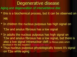
Degenerative changes of the disc Pathological changes • Water and proteoglycan content decreases • Collagen fibers of AF become distorted • Tears may occur in the lamellae • Results in: • Decreased disc height and volume • Decreased resistance to loads
Risk factors Increasing age; Heredity plays an important role; Twin studies revealing similar incidence despite different occupations, socioeconomic status Smoking; Occupation/leisure activity likely does not play a major role; Body habitus;
Pathophysiology Decreased water content in nucleus pulposus • Causes loss of disc height, causing facet joints to override each other; • Facet joints respond with hypertrophy and osteophyte formation; • Can lead to compression of neurological structures, and/or to abnormal movement which worsens the cycle;
Degenerative changes of the vertebral body • Sclerosis: Increased bone formation at the endplates • Reduced nutrition supply • Reduced ability to absorb loads • Osteophytes: Formation of small bony spurs
Degenerative changes of the facet joint Degenerative Changes • Cartilage lining loses water content • Cartilage wears away • Facets override each other • Leads to abnormal function of motion segment
Degenerative changes of the ligaments • Degenerative Changes • Partial ruptures, necrosis and calcifications • Negatively impact function of motion segment
Clinical implications • Axial pain – neck or back • Due to inflammation surrounding diseased structures or to instability of the spine • Neurologic compression • Compresses laterally to nerve root • Radiculopathy • Compresses centrally in canal • In cervical spine: myelopathy • In lumbar spine: neurogenic claudication or cauda equina syndrome
Back pain • 80% adults will have episode back pain; • Most improve over time, therefore initial rest period (short) followed by early mobilization, PT, NSAIDS, lifestyle modification is the treatment; • 90% are not associated with specific discernable cause! (Idiopathic back pain);
Back pain • Red flags (fevers, night sweats, neurological symptoms, weight loss, cancer), severe pain not improving warrant further imaging. • Guidelines published on when to image, types of conservative treatment • Xray, MRI
Radiculopathy Arm pain; leg pain, sciatica; Due to compression lateral to the spinal cord in cervical spine, distal or lateral to nerver root/cauda equina in lumbar spine; Thoracic radiculopathy rare Most common is C5/6, then C6/7; In L spine most common is L5/S1 then L4/5;
Radiculopathy – clinical Pain is the most prominent, along dermatome of affected root;
Lumbar disc herniation • With disruption of the anulus, the soft nucleus was pushed through (herniated) the annulus. • Herniation occurs through a tear in the anulusfibrosus. • Most common at L4/5 and L5/S1 levels, and then L3/4 level; • Herniated disc at upper L spine is rare.
Pathoanatomy • Paracentral herniation is most common; • Paracentral herniation tends to affect nerve root of one level lower! L3/4 DH: affects L4 root; L4/5 DH: affects L5 root; L5/S1 DH: affects S1 root;
LDH and Sciatica • The most classic symptom of a herniated disc is radicular pain in the lower extremity following a dermatomal distribution: sciatica. • Mechanical compression; • Neuroischemia-->inflammation; • Neurochemical factors: immune response • Focal neurologic deficits;
LDH and back pain • Most patients with symptomatic disc herniations present with leg and back pain. • The disc is almost aneural, so where is the pain from? • Mechanical alternation? Innervation of a long degenerated disc? Biochemical irritation?
Classification of LDH Extruded Sequestered Protrusions
History and symptoms • long-standing mild to moderate back pain; • May have a specific incident attributable to the onset of leg and back pain; • Axial back pain is typically present; • Buttock pain: can be referred or radicular in nature • Radicular pain is more typical and often the more “treatable” of the complaints;
Patterns of radiculopathy • S1 radicular pain may radiate to the back of the calf or the lateral aspect or sole of the foot; • L5 radicular pain can lead to symptoms on the dorsum of the foot; • L4 radiculopathy: above or below the knee; • L2 and L3 radiculopathy can produce anterior or medial thigh and groin pain
Physical Examinations • Inspection: • Abnormal gait: limping, slapping; footdrop; • Alignment of the spine Extension: loss of lumbar lordosis, scoliosis; • Palpation and Percussion: • Tenderness at multiple levels; • Local percussion; • Paraspinal muscle spasm;
Neurologic Examination (1) • Sensation: (normal, diminished, or absent ) • L4 sensory function is tested at the medial ankle; • L5 at the first webspace between the great and second toes; • S1 at the lateral aspect of the sole of the foot;
Neurologic Examination (2) • Motor examination • L4 involvement most often affects ankle dorsiflexion (anterior tibialis); • L5 is tested by toe dorsiflexion, particularly the great toe (extensor hallucis longus), and hip abduction. • S1 motor function is assessed by testing plantar flexion;
Neurologic Examination (3) • Deep tendon reflexes • The patellar tendon reflex may be diminished or absent with L3 or L4 involvement; • The Achilles tendon reflex is affected primarily by S1; • There is no specific reflex that reliably reflects L5 function.
Specific tests • Straight leg raising test (SLT): reproduce sciatica at 35-70 degrees; (for L4, L5 & S1 radiculopathy); • Lasègue maneuver; • The femoral stretch test: reproduce anterior thigh pain (for upper root pathology);
Imaging • X-ray: show spinal degenerative changes but not a herniated disc; rule out obvious underlying problems; • CT: relatively less used; • MRI: The best;
Differential diagnosis • The differential diagnosis should be narrowed based on history, physical examination, and selected imaging tests. • idiopathic low back pain; sprain or strain; • spinal stenosis; • Abscess; tuberculosis; • Tumor; • Intrinsic nerve problems;
Nonoperative Treatment • Physiotherapy: Bed rest should be limited to no more than 2 to 3 days; restore strength, flexibility, and function; • Pharmacologic Treatment: Nonsteroidal anti-inflammatory drugs (NSAIDs) are first-line agents; muscle relaxants; • Selective transforaminal steroid injections;
Natural History • A benign disease: Saal and Saal a 90% good or excellent outcome in patients treated nonoperatively; • Another study: at 1 year, 33% had good results, 49% had a fair result, and 18% had a poor result. At 4 years, good results were reported in 51%, fair results were reported in 39%, and poor or bad results were reported in 10%. • 10-year follow-up results: 61% improvement in the predominant symptom, 40% resolution of low back symptoms, and 56% satisfaction rate.
Operative Treatment • Indications • progressive neurologic deficit; • cauda equina syndrome; • failure of appropriate nonoperative treatment;
Discectomy Release ligamentum Inspect neural foramen Remove disc tissues Resect lamina
Cervical spondylosis • Cervical discs similar to lumbar discs, but: • Nucleus pulpous smaller • Discs better supported on lateral margins • Most cervical disc herniations occur in postero-lateral margins
Cervical disc herniation • Patients usually present with one or more of: • Axial neck pain • Radicular arm pain • Myelopathy • Neurapraxia of upper extremities • Non-specific symptoms: dizzying, nausea, head ache, upper back pain;
Treatment of radiculopathy • Nonoperative Treatment • Cervical radiculopathy often resolves without surgery • Conservative methods include PT and anti-inflammatory medicines • Indications for surgery • Continued pain or progressive neurological deficit indicate need for surgery • Anterior and posterior approaches may be used • Fusion with or without instrumentation may be done
Typical surgery: ACDF Anterior cervical decompression and fusion (ACDF); Anterior discectomy; Bone graft or cage; Instrumentation;
Myelopathy (1) A group of symptoms resulting from spinal cord compression, including: • Hand dysfunction • Distal often more affected • Difficulty with buttons, handwriting • Otherwise, extensor pattern ‘pyramidal pattern’ • Triceps, wrist extension • Leg dysfunction • Balance difficulty • Staggering gait • Tandem gait difficulty very early finding
Myelopathy (2) • Sensory disturbance • Often bilateral hand difficulty, sensory level as disease is more severeait • Upper motor neuron signs • Babinski response, hyperreflexia, Hoffman’s sign, increased tone, stiff gait
Degenerative myelopathy – natural history • Typically that of worsening; • Stepwise in 50%, progressive in 50%; • Therefore, patients with myelopathy are usually treated surgically; • Surgery typically performed in expedited fashion; • Relative to rate of deterioration • Lost neurological function is often not regained – the reason to perform early surgery
Surgery Laminaplasty Laminectomy
Lumbar spine stenosis (LSS) • A narrowing of the spinal canal; • one of the most common conditions in the elderly; • Can occur in asymptomatic individuals: Radiographic stenosis is common; • in adults older than 65, LSS is the most common reason to undergo lumbar spine surgery;
Three shapes of the spinal canal The narrowed canal