Download
1 / 36
380 likes | 445 Views
Transhepatic venous cardiac catheterization. David Shim, MD Division of Pediatric Cardiology The Heart Center Children's Hospital Medical Center Cincinnati, Ohio. Indications for right heart catheterization. Hemodynamics right heart pressures pulmonary vascular resistance

E N D
Transhepatic venous cardiac catheterization David Shim, MD Division of Pediatric Cardiology The Heart Center Children's Hospital Medical Center Cincinnati, Ohio
Indications for right heart catheterization • Hemodynamics • right heart pressures • pulmonary vascular resistance • thermodilution cardiac output • Angiography • right ventricular function • pulmonary valve and artery anatomy
Indications for right heart catheterization • Electrophysiology • radiofrequency ablation • Interventions • ASD occlusion • balloon atrial septostomy • endomyocardial biopsy • prograde PDA coil embolization • pulmonary artery balloon dilation/stent
Indications for right heart catheterization • Interventions (continued) • pulmonary valve balloon dilation • RV-PA conduit balloon dilation/stent • SVC balloon dilation/stent • transseptal puncture
Reasons for no access • previous central lines or catheterization • interrupted inferior vena cava • obstructed superior vena cava • bidirectional Glenn/Hemifontan • infection at site of access • devices (eg, Greenfield filter)
Background • Percutaneous Transhepatic Cholangiography (PTC) • has been performed for 2 decades with low morbidity • other transhepatic procedures • portal venous system hemodynamics • localize occult neuroendocrine tumors • embolization of varices
Contraindications • Abnormal clotting/prothrombin time • Active liver disease or peritonitis • Abnormally draining hepatic veins
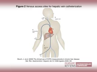
Transhepatic technique • 22 gauge Chiba needle inserted to midlliver under fluoroscopic guidance • needle withdrawn with small injections of contrast until hepatic vein identified • 0.018” Cope wire advanced to RA • 4F coaxial dilator placed and wire exchanged for a 0.035-0.038” guidewire
Transhepatic technique (continued) • dilator removed and curved sheath placed • cardiac catheterization performed • Gianturco coil placed in liver parenchyma upon removal of sheath • puncture site dressed with opsite dressing and post-catheterization care as routine
Evaluation of Efficacy and Safety Shim D, et al. Circulation 1995;92:1526-1530 • Patient population (N=42)
Diagnoses • univentricular heart (25) • critical pulmonary stenosis (5) • tetralogy of Fallot (3) • AV canal (2) • One each: • atrial septal defect, mitral stenosis, • peripheral pulmonary stenosis, • Shone’s complex, status post transplant, • transposition of the great arteries, • and truncus arteriosus
Limitations to access • bilateral femoral venous occlusion (30) • bidirectional Glenn/Hemifontan (9) • interrupted inferior vena cava (7) • obstructed superior vena cava (4) • preferred route for intervention (3) • Greenfield filter (1)
Safety (continued) • Chest radiographs • no effusions • no pneumoperitoneum/pneumothorax • Liver ultrasound (n=34) • small amount of peritoneal fluid (n=4) • no subcapsular hematoma • Clinical hemorrhage (n=2; 5%)
29/30 (97%) successful interventions • angioplasty ± stent • pulmonary (10) • Fontan baffle (3) • superior vena cava (2) • valvuloplasty • pulmonary valve (2) • transseptal mitral valve (1) • radiofrequency ablation • ± transseptal puncture (4) Shim D,et al. Cathet Cardiovasc Interv 1999;47:41-5
Transhepatic interventions • Others • atrial septal defect device occlusion (2) • Fontan fenestration device occlusion (2) • coil embolization of pulmonary artery pseudoaneurysm(2) • device retrieval (1) • endomyocardial biopsy (1) • Sheath sizes: 4-14 French
Conclusions • The transhepatic approachis effective as a route for right sided cardiac catheterization and can be performed with relative safety • The transhepatic approachwill allow therapeutic procedures to be performed in a subset of children where this has been previously not possible
Speculations • Transhepatic access will allow larger sheaths to be used in smaller patients • The transhepatic approach may allow better sheath stability in the right ventricular outflow tract for pulmonary valvuloplasty and angioplasty • The transhepatic approach may also allow a more perpendicular approach to the atrial septum