Download
1 / 20
200 likes | 211 Views
Clinical virology I & II (J12+13). Ondřej Zahradníček Practical sessions of medical microbiology (VLLM0421c) Contact 777 031 969 zahradnicek@fnusa.cz ICQ 242-234-100. Methods of medical microbiology Bold: methods used often in virology. Direct methods (work with a sample or a strain )

E N D
Clinical virology I & II (J12+13) • Ondřej Zahradníček • Practical sessions of medical microbiology (VLLM0421c) • Contact • 777 031 969 zahradnicek@fnusa.cz • ICQ 242-234-100
Methods of medical microbiologyBold: methods used often in virology • Direct methods(work with a sample or a strain) • Microscopy (wet mount, staining…) • Culture (liquid and solid media) • Biochemical and similar identification • Antigen detection (using antibody) • Animal experiment (isolation, toxins) • Nucleic acid detection • Indirect methods • Antibody detection (using antigen)
Direct detection of viruses • Culture isolation (virus is often not multiplied, only kept living). Needs cells. • Microscopy: elektronoptical, optical for detection of in vivo/in vitro effect of viruses (inclusions, cytopathic effects) • Biochemical identification not here • Animal experiment here = viral isolation • DNA/RNA detection – viruses > bacteria • AG detection in sample – common
Indirect detection of viruses • Mostly used: CFT, various neutralisations (HIT, VNT) and in recent time mostly reactions with labelled components (mostly ELISA) • Attention! Not all reaction where serum is used as specimen, are indirect detection methods! In systhemic viroses very often the virus itself or its antigen is present in serum and it is possible to find it here by a direct detection
Viral isolation • Animal is used less often now. Classical animal is a suckling baby-mouse. • Fertilised egg is a classical method: • Amniotic sac • Allantois • Yolk sac • Chorioalantoic membrane (only here sometimes a visible results – tzv. pocs) • Tissue cultures: HeLa, monkey kidney cells, embryonal fibroblasts etc. Some viruses perform a cytopathic effect on tissue cultures.
How can I see a virus, when it is no way to see it? • Bacteria, being cultured, form visible colonies / turbidity. Unlike that, viral isolation visible effects (CPE, pocs) are rare. Usually we have nothing to see. • Detection of an isolated viruses should be performed, e. g. by antigen detection • Most common is Hirst test – detection of viral ability to agglutinate RBC‘s
Microscopy in virology • Elektronoptic microscopy is suitable for observation of majority of viruses, but it is very expensive and not available enough • Optical microscopy may be used • To observe large viruses (poxviruses) • To observe cellullar inclusions in vivo (Negri bodies in rabies/lyssa) • To observe cytopathic effects in vitro (various viruses)
Task 1: ink („virus“) application to amnioric sac • Use ovoscop to see the air membrane • Cut the shell next to air membrane • Apply alcohol to paper membrane • Use your ovoscop again • Usint nidle, try to „stick the egg of the embryo“ (it runs away anyway) • Apply ink inside • Carefully remove the inner structures of the egg to a Petri dish and draw them.
More tasks • Task 2: Put all the test-tubes to the microscope, try to focus to the inner wall of the test tube. Maybe you‘ll see the tissue culture, maybe even a CPE. • Task 3: Do according to instructions. In positive case, you would see agglutinated red blood cells. • Task 4: Perform indirect diagnostics of tick-borne encefalitis virus using HIT • Task 5: Perform indirect diagnostics of several viruses using CFT
Remember! (To tasks 4 & 5) • In HIT, blocation of viral agglutination is positive. Agglutination is negative! • In CFT, absence of haemolysis is positive, haemolysis is negative • Titer = highest serum dilution with still positive reaction • Fourfold increase/decrease of titer is supposed to be significant for running infection when using pair sera
Task No. 6 – shell-vials techniques (demonstration) • Those are techniques of quick culture. They are very popular in last time. It is a direct diatgnostic, but not so long during and complicated like classical viral isolation.
J13: Hepatitis and HIV • There are five main types of viral hepatitis (VHA – VHE) caused by viruses HAV – HEV. They belong to different groups of viruses. HBV is DNA virus, the other are RNA viruses. • VHA and VHE are transmitted by faecal – oral route (hands) • VHB, VHC and VHD – blood and sexual transmission, (sexual – not for VHC) • Virus HIV causes AIDS and two its pre-stages (PGL, ARC)
Task 1 – diagnostics of HAV • Task 1a) is determination of anti-HAV IgM. Counting cut off: average C1 / D1 • Task 1b) is determination of total anti-HAV antibodies (instead of IgG) • Counting cut off (1a + b): • Average of „c. o.“ wells C1 and D1 = cut off • Values 110 % of cut off and more = positive • Values 90 % of cut off and less = negative • Values 90 – 100 % of cut off = borderline
HAV HAV! HAV!
Task 2: HBV diagnostics • HBV has three angigens important for diagnostics. Only two of them are present in serum: HBsAg (common, produced more than the others) and HBeAg. • Antibodies against all three of them may be detected: anti-HBs, anti-HBe and anti-HBc. • Diagnostics may be completed by PCR, liver enzymes etc. • Interpretation is complex
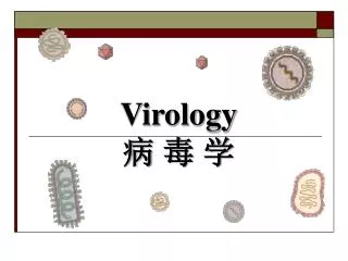
Virus of hepatitis B Only HBsAg 22 nm Complete virion (Dane particle) 42 nm HBsAg, inside it delta agens (VHD) 35 nm
Determination of HBV markers • 2a) Determination of HBsAg a HBeAg • 2b) Determination of anti-HBs, anti-HBe • Average of „c. o.“ wells C1 and D1 = cut off • Values 110 % of cut off and more = positive • Values 90 % of cut off and less = negative • Values 90 – 100 % of cut off = borderline
Task 3: HCV diagnostics • 3a): PCR, elektroforesis • 3b): anti-HCV • Average of „negative control“ wells B1, C1 and D1 + 0.050 = cut off • Values 110 % of cut off and more = positive • Values 90 % of cut off and less = negative • Values 90 – 100 % of cut off = borderline
Task 4: ELISA anti-HIV ELISA antibodies – anti HIV • Average of „c. o.“ wells C1 and D1 = cut off • Values 110 % of cut off and more = positive • Values 90 % of cut off and less = negative • Values 90 – 100 % of cut off = borderline