Download

1 / 45
550 likes | 1.47k Views
The anatomy of the rectum and anus. The muscular arrangement of the levator ani muscles. Factors necessary for maintenance of fecal continence. Triple loop mechanism of external anal sphincter. Introduction. Common medical problem that is under-reported to physiciansSecond leading cause of nursing home placement, more common than dementiaSome degree of fecal incontinence will develop in 3% of women who give birth by vaginal delivery.

E N D
1. Fecal incontinence Tova Rainis
Gastroenterology unit
Bnai-zion
2. Figure 1-38. The anatomy of the rectum and anus is shown. The most distal 7 to 8 cm of rectum is infraperitoneal and is thereby fixed by surrounding supporting tissues. More orad regions of the rectum and sigmoid colons are intraperitoneal and more mobile. The rectal mucosa is smoother and less corrugated than the more proximal colon, and the rectum is capacious owing to its physiologic function as a storage receptacle for feces before defecation. The rectal mucosa forms three eccentric rectal valves, also known as the valves of Houston. The most distal rectum is marked by the rectal columns of Morgagni, which terminate at the pectinate or dentate line delineating the junction of the rectal and anal mucosae. The transition from columnar to squamous mucosa occurs gradually in the distal rectum, usually above the pectinate line. The hemorrhoidal plexuses reside under the mucosa of both the rectum and anus and are believed to serve a cushioning role. The anal musculature is specialized with the circular muscle layer forming a prominent internal anal sphincter. The longitudinal muscle of the rectum projects to the region of the internal anal sphincter but is not as prominent. The external anal sphincter, a skeletal muscle structure, is composed of three muscle groups, the deep external sphincter, the superficial external sphincter, and the subcutaneous external sphincter. The internal sphincter is responsible for much of the resting anal tone. The external sphincter responds to voluntary squeeze commands to maintain continence when feces have moved into the rectum. Finally, the levator ani muscles, three skeletal muscles (puborectalis, pubococcygeus, and iliococcygeus), form the sling musculature surrounding the distal rectum that maintain continence when contracted. When the puborectalis component of the levator ani apparatus relaxes, defecation is permitted.Figure 1-38. The anatomy of the rectum and anus is shown. The most distal 7 to 8 cm of rectum is infraperitoneal and is thereby fixed by surrounding supporting tissues. More orad regions of the rectum and sigmoid colons are intraperitoneal and more mobile. The rectal mucosa is smoother and less corrugated than the more proximal colon, and the rectum is capacious owing to its physiologic function as a storage receptacle for feces before defecation. The rectal mucosa forms three eccentric rectal valves, also known as the valves of Houston. The most distal rectum is marked by the rectal columns of Morgagni, which terminate at the pectinate or dentate line delineating the junction of the rectal and anal mucosae. The transition from columnar to squamous mucosa occurs gradually in the distal rectum, usually above the pectinate line. The hemorrhoidal plexuses reside under the mucosa of both the rectum and anus and are believed to serve a cushioning role. The anal musculature is specialized with the circular muscle layer forming a prominent internal anal sphincter. The longitudinal muscle of the rectum projects to the region of the internal anal sphincter but is not as prominent. The external anal sphincter, a skeletal muscle structure, is composed of three muscle groups, the deep external sphincter, the superficial external sphincter, and the subcutaneous external sphincter. The internal sphincter is responsible for much of the resting anal tone. The external sphincter responds to voluntary squeeze commands to maintain continence when feces have moved into the rectum. Finally, the levator ani muscles, three skeletal muscles (puborectalis, pubococcygeus, and iliococcygeus), form the sling musculature surrounding the distal rectum that maintain continence when contracted. When the puborectalis component of the levator ani apparatus relaxes, defecation is permitted.
3. Figure 1-39. The muscular arrangement of the levator ani muscles is shown in relation to the anorectum and pelvis. Some of the levator ani muscles merge with fibers of the external anal sphincter. Each of the levator muscles has a bony attachment on the anterior of the pelvis. Only the iliococcygeus is fixed posteriorly to the coccyx via the anococcygeal raphe. The puborectalis is tonically contracted under resting conditions, pinching closed the distal rectum and aiding the anal sphincter in maintenance of continence. With defecation, the puborectalis component of the levator ani muscles relaxes and permits expulsion of the fecal bolus. (Adapted from Mulholland [2]; with permission.)Figure 1-39. The muscular arrangement of the levator ani muscles is shown in relation to the anorectum and pelvis. Some of the levator ani muscles merge with fibers of the external anal sphincter. Each of the levator muscles has a bony attachment on the anterior of the pelvis. Only the iliococcygeus is fixed posteriorly to the coccyx via the anococcygeal raphe. The puborectalis is tonically contracted under resting conditions, pinching closed the distal rectum and aiding the anal sphincter in maintenance of continence. With defecation, the puborectalis component of the levator ani muscles relaxes and permits expulsion of the fecal bolus. (Adapted from Mulholland [2]; with permission.)
4. Figure 1-42. The factors necessary for maintenance of fecal continence are shown. During its passage through the colon, fecal material becomes dehydrated and formed such that the stool that reaches the rectum is semisolid to solid. The rectum itself exhibits a highly compliant wall that allows it to serve as a reservoir for the fecal material until it can be conveniently expelled. The internal anal sphincter provides sufficient tone to prevent accidental loss of fecal material, whereas the external sphincter muscles voluntarily contract if unwanted loss of feces is impending. The anal cushions, made up of mucosa, hemorrhoidal plexuses, and the subepithelial-supporting tissue provide a seal that does not permit passage of liquid material under resting conditions. Finally, the anorectal angle created by the puborectalis muscle provides a functional obstruction to accidental loss of stool at rest. (Adapted from Irving and Catchpole [30]; with permission.)Figure 1-42. The factors necessary for maintenance of fecal continence are shown. During its passage through the colon, fecal material becomes dehydrated and formed such that the stool that reaches the rectum is semisolid to solid. The rectum itself exhibits a highly compliant wall that allows it to serve as a reservoir for the fecal material until it can be conveniently expelled. The internal anal sphincter provides sufficient tone to prevent accidental loss of fecal material, whereas the external sphincter muscles voluntarily contract if unwanted loss of feces is impending. The anal cushions, made up of mucosa, hemorrhoidal plexuses, and the subepithelial-supporting tissue provide a seal that does not permit passage of liquid material under resting conditions. Finally, the anorectal angle created by the puborectalis muscle provides a functional obstruction to accidental loss of stool at rest. (Adapted from Irving and Catchpole [30]; with permission.)
5. Figure 1-44. The external anal sphincter assists in the maintenance of fecal continence by way of the triple loop mechanism shown in this figure. On the left, the three loops are formed by the three distinct muscles of the external anal sphincter. The uppermost loop, formed from the deep external sphincter muscle, attaches anteriorly on the pubis. The intermediate loop, formed from the superficial external sphincter, attaches posteriorly to the coccyx. The base loop, formed from the subcutaneous external sphincter, attaches to the perianal skin. On the right, a diagrammatic representation of the mechanism of anal occlusion by the triple loop system of the external anal sphincter is shown. A, The external sphincter at rest. With external sphincter contraction (B), the direction of contraction of each of the loops is shown (anterior to the left, posterior to the right). Tight contraction of these three loops results in tight anal occlusion as shown in C. (Adapted from Shafik [31] and Shafik [32]; with permission.)Figure 1-44. The external anal sphincter assists in the maintenance of fecal continence by way of the triple loop mechanism shown in this figure. On the left, the three loops are formed by the three distinct muscles of the external anal sphincter. The uppermost loop, formed from the deep external sphincter muscle, attaches anteriorly on the pubis. The intermediate loop, formed from the superficial external sphincter, attaches posteriorly to the coccyx. The base loop, formed from the subcutaneous external sphincter, attaches to the perianal skin. On the right, a diagrammatic representation of the mechanism of anal occlusion by the triple loop system of the external anal sphincter is shown. A, The external sphincter at rest. With external sphincter contraction (B), the direction of contraction of each of the loops is shown (anterior to the left, posterior to the right). Tight contraction of these three loops results in tight anal occlusion as shown in C. (Adapted from Shafik [31] and Shafik [32]; with permission.)
6. Introduction Common medical problem that is under-reported to physicians
Second leading cause of nursing home placement, more common than dementia
Some degree of fecal incontinence will develop in 3% of women who give birth by vaginal delivery
7. Pathophysiology and etiology Partial incontinence � loss of control to flatus and minor soiling
Major incontinence � frequent and regular deficiency in the ability to control stool of normal consistency
8. Four basic physiologic factors:
stool consistency,
rectal compliance,
rectal and anal sensation
pelvic floor function
can lead to a defective continence mechanism
9. Incontinence with normal pelvic floor function Altered stool consistency
Inflammatory bowel disease
Infectious diarrhea
Laxative abuse
Radiation enteritis
Short bowel syndrome
Malabsorption syndrome
10. Incontinence with normal pelvic floor function - 2 Inadequate rectal compliance
Inflammatory bowel disease
Absent rectal reservoir (ileoanal, low ant. resection)
Rectal ischemia
Collagen vascular disease (scleroderma, amyloidosis, dermatomyositis)
Rectal neoplasms
11. Incontinence with normal pelvic floor function -3 Inadequate rectal sensation
Dementia, CVA, MS, brain or spinal cord injury/neoplasm, sensory neuropathy, tabes dorsalis
Overflow incontinence
Fecal impaction � leading cause of incontinence in institutionalized elderly patients
Diabetes � multifactorial, impaired rectal sensation is important
12. Figure 1-47. The reflex responsiveness of the anal region to a distending stimulus in the rectum is shown. Rectal balloon inflation results in an abrupt decrease in pressure in the internal anal sphincter, known as the rectoanal inhibitory reflex (top). In contrast, there is a normal increase in pressure in the external anal sphincter upon rectal distention (bottom). These physiologic responses are important for two reasons. The presence of a rectoanal inhibitory reflex allows the anus to relax upon filling of the rectal vault to permit efficient defecation. The reflex contraction of the external sphincter prevents accidental leakage of rectal contents until they may be evacuated in a socially convenient setting. (Adapted from Alva et al. [35]; with permission.)Figure 1-47. The reflex responsiveness of the anal region to a distending stimulus in the rectum is shown. Rectal balloon inflation results in an abrupt decrease in pressure in the internal anal sphincter, known as the rectoanal inhibitory reflex (top). In contrast, there is a normal increase in pressure in the external anal sphincter upon rectal distention (bottom). These physiologic responses are important for two reasons. The presence of a rectoanal inhibitory reflex allows the anus to relax upon filling of the rectal vault to permit efficient defecation. The reflex contraction of the external sphincter prevents accidental leakage of rectal contents until they may be evacuated in a socially convenient setting. (Adapted from Alva et al. [35]; with permission.)
13. Figure 5-6. Fecal incontinence associated with spinal cord injury. In a manometric study of a normal subject, rectal balloon distension (arrow) results in transient external anal sphincter contraction. In a patient with meningomyelocele and sacral denervation, rectal balloon distention (arrow) elicits normal internal anal sphincter relaxation, but there is no contraction response from the external anal sphincter; therefore, decreased pressures in the anal canal are recorded by the external anal sphincter balloon. Rectal sensation in these patients may be normal or impaired.Figure 5-6. Fecal incontinence associated with spinal cord injury. In a manometric study of a normal subject, rectal balloon distension (arrow) results in transient external anal sphincter contraction. In a patient with meningomyelocele and sacral denervation, rectal balloon distention (arrow) elicits normal internal anal sphincter relaxation, but there is no contraction response from the external anal sphincter; therefore, decreased pressures in the anal canal are recorded by the external anal sphincter balloon. Rectal sensation in these patients may be normal or impaired.
14. Incontinence with abnormal pelvic floor function Anatomic sphincter defect � internal or external
Traumatic
Obstetric injury � prolonged difficult labor with forceps application, episiotomy complications, third or fourth-degree lacerations
Anorectal surgery � anal fistula surgery - most common operative procedure that results in fecal incontinence; hemorrhoidectomy
15. Incontinence with abnormal pelvic floor function - 2 Pelvic floor denervation � degenerative neurogenic factors are a common cause of non-surgically related incontinence
Primary (idiopathic neurogenic incontinence)
Pudendal neuropathy � 80%. Denervation of the puborectalis muscle and external anal sphincter muscles � results in an impaired ability to maintain the anorectal angle and prevent gross incontinence
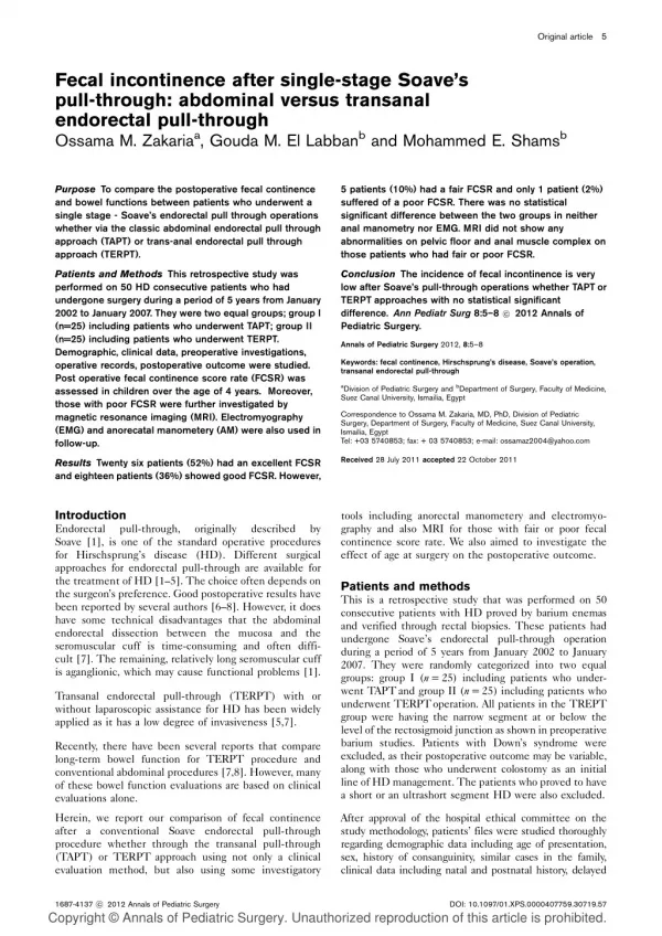
16. Figure 5-8. Fecal incontinence associated with pudendal neuropathy. A and B, Barium proctography in a normal subject. With the patient in the left lateral recumbent position, 200 to 250 mL of barium contrast medium is instilled to fill the rectum and distal sigmoid colon. A small plastic lubricated sphere attached to a chain of radiopaque beads is inserted into the anal canal, and the chain is taped to the dorsal intergluteal cleft. Lateral radiographs are obtained at rest, when the patient squeezes, and when the patient strains while attempting to retain the barium. The anorectal angle is calculated from the intersection of a line drawn along the posterior rectal wall with a line drawn along the axis of the anal canal, as delineated by the beaded chain. The position of the pelvic floor is determined by the junction of the anorectal angle and compared with its normal position which approximates a line drawn from the symphysis pubis to the tip of the coccyx. This technique has been superseded by defecating proctography, in which 200 mL of thickened barium paste is administered into the rectum using a wide-tipped syringe. The paste consists of barium contrast mixed with a thickening agent such as hydrophilic psyllium colloid. This technique eliminates the need for a chain to outline the anal canal.C and D, Fecal incontinence with perineal descent. In contrast to a normal subject, the anorectal angle in a patient with fecal incontinence is more obtuse and lies significantly below the pubococcygeal line at rest (C). When the patient strains (D), perineal descent is grossly apparent and there is loss of contrast material through a weakened anal sphincter musculature. (A and B,Courtesy of N.W. Read, Sheffield, UK)Figure 5-8. Fecal incontinence associated with pudendal neuropathy. A and B, Barium proctography in a normal subject. With the patient in the left lateral recumbent position, 200 to 250 mL of barium contrast medium is instilled to fill the rectum and distal sigmoid colon. A small plastic lubricated sphere attached to a chain of radiopaque beads is inserted into the anal canal, and the chain is taped to the dorsal intergluteal cleft. Lateral radiographs are obtained at rest, when the patient squeezes, and when the patient strains while attempting to retain the barium. The anorectal angle is calculated from the intersection of a line drawn along the posterior rectal wall with a line drawn along the axis of the anal canal, as delineated by the beaded chain. The position of the pelvic floor is determined by the junction of the anorectal angle and compared with its normal position which approximates a line drawn from the symphysis pubis to the tip of the coccyx. This technique has been superseded by defecating proctography, in which 200 mL of thickened barium paste is administered into the rectum using a wide-tipped syringe. The paste consists of barium contrast mixed with a thickening agent such as hydrophilic psyllium colloid. This technique eliminates the need for a chain to outline the anal canal.C and D, Fecal incontinence with perineal descent. In contrast to a normal subject, the anorectal angle in a patient with fecal incontinence is more obtuse and lies significantly below the pubococcygeal line at rest (C). When the patient strains (D), perineal descent is grossly apparent and there is loss of contrast material through a weakened anal sphincter musculature. (A and B,Courtesy of N.W. Read, Sheffield, UK)
17. Figure 5-8. Fecal incontinence associated with pudendal neuropathy. A and B, Barium proctography in a normal subject. With the patient in the left lateral recumbent position, 200 to 250 mL of barium contrast medium is instilled to fill the rectum and distal sigmoid colon. A small plastic lubricated sphere attached to a chain of radiopaque beads is inserted into the anal canal, and the chain is taped to the dorsal intergluteal cleft. Lateral radiographs are obtained at rest, when the patient squeezes, and when the patient strains while attempting to retain the barium. The anorectal angle is calculated from the intersection of a line drawn along the posterior rectal wall with a line drawn along the axis of the anal canal, as delineated by the beaded chain. The position of the pelvic floor is determined by the junction of the anorectal angle and compared with its normal position which approximates a line drawn from the symphysis pubis to the tip of the coccyx. This technique has been superseded by defecating proctography, in which 200 mL of thickened barium paste is administered into the rectum using a wide-tipped syringe. The paste consists of barium contrast mixed with a thickening agent such as hydrophilic psyllium colloid. This technique eliminates the need for a chain to outline the anal canal.C and D, Fecal incontinence with perineal descent. In contrast to a normal subject, the anorectal angle in a patient with fecal incontinence is more obtuse and lies significantly below the pubococcygeal line at rest (C). When the patient strains (D), perineal descent is grossly apparent and there is loss of contrast material through a weakened anal sphincter musculature. (A and B,Courtesy of N.W. Read, Sheffield, UK)Figure 5-8. Fecal incontinence associated with pudendal neuropathy. A and B, Barium proctography in a normal subject. With the patient in the left lateral recumbent position, 200 to 250 mL of barium contrast medium is instilled to fill the rectum and distal sigmoid colon. A small plastic lubricated sphere attached to a chain of radiopaque beads is inserted into the anal canal, and the chain is taped to the dorsal intergluteal cleft. Lateral radiographs are obtained at rest, when the patient squeezes, and when the patient strains while attempting to retain the barium. The anorectal angle is calculated from the intersection of a line drawn along the posterior rectal wall with a line drawn along the axis of the anal canal, as delineated by the beaded chain. The position of the pelvic floor is determined by the junction of the anorectal angle and compared with its normal position which approximates a line drawn from the symphysis pubis to the tip of the coccyx. This technique has been superseded by defecating proctography, in which 200 mL of thickened barium paste is administered into the rectum using a wide-tipped syringe. The paste consists of barium contrast mixed with a thickening agent such as hydrophilic psyllium colloid. This technique eliminates the need for a chain to outline the anal canal.C and D, Fecal incontinence with perineal descent. In contrast to a normal subject, the anorectal angle in a patient with fecal incontinence is more obtuse and lies significantly below the pubococcygeal line at rest (C). When the patient strains (D), perineal descent is grossly apparent and there is loss of contrast material through a weakened anal sphincter musculature. (A and B,Courtesy of N.W. Read, Sheffield, UK)
18. Figure 5-8. Fecal incontinence associated with pudendal neuropathy. A and B, Barium proctography in a normal subject. With the patient in the left lateral recumbent position, 200 to 250 mL of barium contrast medium is instilled to fill the rectum and distal sigmoid colon. A small plastic lubricated sphere attached to a chain of radiopaque beads is inserted into the anal canal, and the chain is taped to the dorsal intergluteal cleft. Lateral radiographs are obtained at rest, when the patient squeezes, and when the patient strains while attempting to retain the barium. The anorectal angle is calculated from the intersection of a line drawn along the posterior rectal wall with a line drawn along the axis of the anal canal, as delineated by the beaded chain. The position of the pelvic floor is determined by the junction of the anorectal angle and compared with its normal position which approximates a line drawn from the symphysis pubis to the tip of the coccyx. This technique has been superseded by defecating proctography, in which 200 mL of thickened barium paste is administered into the rectum using a wide-tipped syringe. The paste consists of barium contrast mixed with a thickening agent such as hydrophilic psyllium colloid. This technique eliminates the need for a chain to outline the anal canal.C and D, Fecal incontinence with perineal descent. In contrast to a normal subject, the anorectal angle in a patient with fecal incontinence is more obtuse and lies significantly below the pubococcygeal line at rest (C). When the patient strains (D), perineal descent is grossly apparent and there is loss of contrast material through a weakened anal sphincter musculature. (A and B,Courtesy of N.W. Read, Sheffield, UK)Figure 5-8. Fecal incontinence associated with pudendal neuropathy. A and B, Barium proctography in a normal subject. With the patient in the left lateral recumbent position, 200 to 250 mL of barium contrast medium is instilled to fill the rectum and distal sigmoid colon. A small plastic lubricated sphere attached to a chain of radiopaque beads is inserted into the anal canal, and the chain is taped to the dorsal intergluteal cleft. Lateral radiographs are obtained at rest, when the patient squeezes, and when the patient strains while attempting to retain the barium. The anorectal angle is calculated from the intersection of a line drawn along the posterior rectal wall with a line drawn along the axis of the anal canal, as delineated by the beaded chain. The position of the pelvic floor is determined by the junction of the anorectal angle and compared with its normal position which approximates a line drawn from the symphysis pubis to the tip of the coccyx. This technique has been superseded by defecating proctography, in which 200 mL of thickened barium paste is administered into the rectum using a wide-tipped syringe. The paste consists of barium contrast mixed with a thickening agent such as hydrophilic psyllium colloid. This technique eliminates the need for a chain to outline the anal canal.C and D, Fecal incontinence with perineal descent. In contrast to a normal subject, the anorectal angle in a patient with fecal incontinence is more obtuse and lies significantly below the pubococcygeal line at rest (C). When the patient strains (D), perineal descent is grossly apparent and there is loss of contrast material through a weakened anal sphincter musculature. (A and B,Courtesy of N.W. Read, Sheffield, UK)
19. Figure 5-8. Fecal incontinence associated with pudendal neuropathy. A and B, Barium proctography in a normal subject. With the patient in the left lateral recumbent position, 200 to 250 mL of barium contrast medium is instilled to fill the rectum and distal sigmoid colon. A small plastic lubricated sphere attached to a chain of radiopaque beads is inserted into the anal canal, and the chain is taped to the dorsal intergluteal cleft. Lateral radiographs are obtained at rest, when the patient squeezes, and when the patient strains while attempting to retain the barium. The anorectal angle is calculated from the intersection of a line drawn along the posterior rectal wall with a line drawn along the axis of the anal canal, as delineated by the beaded chain. The position of the pelvic floor is determined by the junction of the anorectal angle and compared with its normal position which approximates a line drawn from the symphysis pubis to the tip of the coccyx. This technique has been superseded by defecating proctography, in which 200 mL of thickened barium paste is administered into the rectum using a wide-tipped syringe. The paste consists of barium contrast mixed with a thickening agent such as hydrophilic psyllium colloid. This technique eliminates the need for a chain to outline the anal canal.C and D, Fecal incontinence with perineal descent. In contrast to a normal subject, the anorectal angle in a patient with fecal incontinence is more obtuse and lies significantly below the pubococcygeal line at rest (C). When the patient strains (D), perineal descent is grossly apparent and there is loss of contrast material through a weakened anal sphincter musculature. (A and B,Courtesy of N.W. Read, Sheffield, UK)Figure 5-8. Fecal incontinence associated with pudendal neuropathy. A and B, Barium proctography in a normal subject. With the patient in the left lateral recumbent position, 200 to 250 mL of barium contrast medium is instilled to fill the rectum and distal sigmoid colon. A small plastic lubricated sphere attached to a chain of radiopaque beads is inserted into the anal canal, and the chain is taped to the dorsal intergluteal cleft. Lateral radiographs are obtained at rest, when the patient squeezes, and when the patient strains while attempting to retain the barium. The anorectal angle is calculated from the intersection of a line drawn along the posterior rectal wall with a line drawn along the axis of the anal canal, as delineated by the beaded chain. The position of the pelvic floor is determined by the junction of the anorectal angle and compared with its normal position which approximates a line drawn from the symphysis pubis to the tip of the coccyx. This technique has been superseded by defecating proctography, in which 200 mL of thickened barium paste is administered into the rectum using a wide-tipped syringe. The paste consists of barium contrast mixed with a thickening agent such as hydrophilic psyllium colloid. This technique eliminates the need for a chain to outline the anal canal.C and D, Fecal incontinence with perineal descent. In contrast to a normal subject, the anorectal angle in a patient with fecal incontinence is more obtuse and lies significantly below the pubococcygeal line at rest (C). When the patient strains (D), perineal descent is grossly apparent and there is loss of contrast material through a weakened anal sphincter musculature. (A and B,Courtesy of N.W. Read, Sheffield, UK)
20. Incontinence with abnormal pelvic floor function - 3 �Descending perineal syndrome� � results from constant straining during defecation that causes a traction neuropathy of the nerves running along the pelvic floor muscles. Both the puborectalis and external anal sphincter muscles become denervated
Vaginal deliveries
Secondary
Injuries to spinal cord, cauda equina
Diabetic neuropathy
21. Incontinence with abnormal pelvic floor function - 4 Congenital abnornmalities
Spina bifida
Myelomeningocele
Miscellaneous
Rectal prolapse � 60-70% incontinence.
Aging � anal canal pressure and rectal compliance are decreased
22. Diagnostic evaluation History
Few patients will volunteer the symptom on their own � embarrassment, �chronic diarrhea�
Use terms as �leakage�, �soiling� or �accidents� to facilitate communication
Incontinence grading scale
24. Physical examination Search for hemorrhoids, scars from previous surgery, skin tags, fissures, fistulas
Signs of rectal prolapse or descent of the perineum
�anal wink�
Rectal examination � obtain estimation of resting anal sphincter pressure/external anal sphincter strength; fecal impaction
25. Table 5-3. Diagnostic studies for fecal incontinence. Anorectal function can be studied by a variety of complementary techniques, each providing information concerning the various continence mechanisms. Of these, sigmoidoscopy and anorectal manometry are the most useful and are indicated in most patients; the other studies provide valuable information but are not necessary in all patients.Table 5-3. Diagnostic studies for fecal incontinence. Anorectal function can be studied by a variety of complementary techniques, each providing information concerning the various continence mechanisms. Of these, sigmoidoscopy and anorectal manometry are the most useful and are indicated in most patients; the other studies provide valuable information but are not necessary in all patients.
26. Anorectal manometry Measurement of both resting and voluntary sphincter squeeze pressure
Incontinent patients � low resting and voluntary squeeze pressure
The study cannot discriminate between primary muscle and neuronal defects
Estimate threshold for rectal sensation/compliance, rectoanal inhibitory reflex
27. Figure 5-13. Anorectal manometry in fecal incontinence. A, In many patients with fecal incontinence, external anal sphincter contraction following rectal distension is often absent or occurs only at much higher distending volumes than in normal subjects. In this tracing, rectal distension (arrows) induces relaxation of the internal anal sphincter, but no external sphincter contractions are observed. Reestablishing phasic contractions of the external anal sphincter is one aspect of sphincter muscle retraining using biofeedback techniques. B, Manometric tracing. The manometric tracings of a prolonged voluntary contraction from a healthy volunteer (bottom) is compared with a tracing from a patient with fecal incontinence (top). A rapidly fatiguing anal sphincter during an attempt of prolonged squeezing is illustrated in the patient with fecal incontinence. (A, From Engel et al. [9]; with permission.) (B, From Chiaroni et al. [10]; with permission.)Figure 5-13. Anorectal manometry in fecal incontinence. A, In many patients with fecal incontinence, external anal sphincter contraction following rectal distension is often absent or occurs only at much higher distending volumes than in normal subjects. In this tracing, rectal distension (arrows) induces relaxation of the internal anal sphincter, but no external sphincter contractions are observed. Reestablishing phasic contractions of the external anal sphincter is one aspect of sphincter muscle retraining using biofeedback techniques. B, Manometric tracing. The manometric tracings of a prolonged voluntary contraction from a healthy volunteer (bottom) is compared with a tracing from a patient with fecal incontinence (top). A rapidly fatiguing anal sphincter during an attempt of prolonged squeezing is illustrated in the patient with fecal incontinence. (A, From Engel et al. [9]; with permission.) (B, From Chiaroni et al. [10]; with permission.)
28. Figure 5-13. Anorectal manometry in fecal incontinence. A, In many patients with fecal incontinence, external anal sphincter contraction following rectal distension is often absent or occurs only at much higher distending volumes than in normal subjects. In this tracing, rectal distension (arrows) induces relaxation of the internal anal sphincter, but no external sphincter contractions are observed. Reestablishing phasic contractions of the external anal sphincter is one aspect of sphincter muscle retraining using biofeedback techniques. B, Manometric tracing. The manometric tracings of a prolonged voluntary contraction from a healthy volunteer (bottom) is compared with a tracing from a patient with fecal incontinence (top). A rapidly fatiguing anal sphincter during an attempt of prolonged squeezing is illustrated in the patient with fecal incontinence. (A, From Engel et al. [9]; with permission.) (B, From Chiaroni et al. [10]; with permission.)Figure 5-13. Anorectal manometry in fecal incontinence. A, In many patients with fecal incontinence, external anal sphincter contraction following rectal distension is often absent or occurs only at much higher distending volumes than in normal subjects. In this tracing, rectal distension (arrows) induces relaxation of the internal anal sphincter, but no external sphincter contractions are observed. Reestablishing phasic contractions of the external anal sphincter is one aspect of sphincter muscle retraining using biofeedback techniques. B, Manometric tracing. The manometric tracings of a prolonged voluntary contraction from a healthy volunteer (bottom) is compared with a tracing from a patient with fecal incontinence (top). A rapidly fatiguing anal sphincter during an attempt of prolonged squeezing is illustrated in the patient with fecal incontinence. (A, From Engel et al. [9]; with permission.) (B, From Chiaroni et al. [10]; with permission.)
29. Electrophysiologic tests EMG � needle electrodes into the superficial portion of the external sphincter or puborectalis muscle � myoelectric activity
Pudendal nerve terminal motor latency � measures the delay between the application of an electrical stimulus and external sphincter muscle response. Prolonged � pudendal neuropathy
30. Defecography Videodefecography � barium thickened to the consistency of stool is introduced into the rectum.
Evacuation is monitored with flouroscopy
Assessment of the anorectal angle at rest and during defecation
Excessive perineal descent, failure of the puborectalis muscle to relax, rectocele and internal intususception
31. Figure 5-27. A, Rectocele. During expulsion, a large anterior rectocele fills with barium (arrow), but expulsion of contrast appears normal. Several studies have demonstrated that rectoceles are common in women without defecation complaints so that caution is necessary before implicating a rectocele as the cause of defecation difficulty. B, Rectal intussusception. At left, the anorectum is filled with barium paste and appears normal. During attempted evacuation (right), inversion of the rectal mucosa occurs during maximal straining (arrows). No further emptying occurs in this patient. The vagina (V) is identified by a radiocontrast soaked tampon and lies anterior to the rectum. (A, Courtesy of D. Bauman, Pittsburgh, PA)Figure 5-27. A, Rectocele. During expulsion, a large anterior rectocele fills with barium (arrow), but expulsion of contrast appears normal. Several studies have demonstrated that rectoceles are common in women without defecation complaints so that caution is necessary before implicating a rectocele as the cause of defecation difficulty. B, Rectal intussusception. At left, the anorectum is filled with barium paste and appears normal. During attempted evacuation (right), inversion of the rectal mucosa occurs during maximal straining (arrows). No further emptying occurs in this patient. The vagina (V) is identified by a radiocontrast soaked tampon and lies anterior to the rectum. (A, Courtesy of D. Bauman, Pittsburgh, PA)
32. Anal Endosonography An ultrasound probe is placed in the anal canal or transvaginally to detect sphincter injuries and to evaluate pelvic floor structures
33. Figure 5-12. A, Normal anatomy as viewed by anal endosonography. The cone around the sonographic probe creates two inner bright interfaces. Just outside the interfaces, the subepithelial tissues are seen to be slightly hyperechoic. The internal sphincter creates a well-defined hypoechoic ring (closed arrows). The hyperechoic longitudinal muscle surrounds the internal sphincter, and the external sphincter (open arrows) envelopes the longitudinal muscle. In this male patient, the external sphincter is hypoechoic relative to the longitudinal muscle. In most females, these sections are equally echogenic and cannot be identified separately. The transverse perineii are noted anteriorly (curved arrow). The image is oriented so that anterior is in the uppermost position. B, Lateral internal anal sphincterotomy within the 6 to 10 o'clock positions (arrow). The remaining internal anal sphincter has bunched up slightly and appears somewhat thickened. Surgical defects in the internal anal sphincter are usually clear-cut with well-defined ends. C, Obstetric trauma caused by forceps delivery with internal and external defects within the 9 to 1 o'clock positions (arrows). (Courtesy of C.I. Bartram, London, UK)Figure 5-12. A, Normal anatomy as viewed by anal endosonography. The cone around the sonographic probe creates two inner bright interfaces. Just outside the interfaces, the subepithelial tissues are seen to be slightly hyperechoic. The internal sphincter creates a well-defined hypoechoic ring (closed arrows). The hyperechoic longitudinal muscle surrounds the internal sphincter, and the external sphincter (open arrows) envelopes the longitudinal muscle. In this male patient, the external sphincter is hypoechoic relative to the longitudinal muscle. In most females, these sections are equally echogenic and cannot be identified separately. The transverse perineii are noted anteriorly (curved arrow). The image is oriented so that anterior is in the uppermost position. B, Lateral internal anal sphincterotomy within the 6 to 10 o'clock positions (arrow). The remaining internal anal sphincter has bunched up slightly and appears somewhat thickened. Surgical defects in the internal anal sphincter are usually clear-cut with well-defined ends. C, Obstetric trauma caused by forceps delivery with internal and external defects within the 9 to 1 o'clock positions (arrows). (Courtesy of C.I. Bartram, London, UK)
34. Figure 5-12. A, Normal anatomy as viewed by anal endosonography. The cone around the sonographic probe creates two inner bright interfaces. Just outside the interfaces, the subepithelial tissues are seen to be slightly hyperechoic. The internal sphincter creates a well-defined hypoechoic ring (closed arrows). The hyperechoic longitudinal muscle surrounds the internal sphincter, and the external sphincter (open arrows) envelopes the longitudinal muscle. In this male patient, the external sphincter is hypoechoic relative to the longitudinal muscle. In most females, these sections are equally echogenic and cannot be identified separately. The transverse perineii are noted anteriorly (curved arrow). The image is oriented so that anterior is in the uppermost position. B, Lateral internal anal sphincterotomy within the 6 to 10 o'clock positions (arrow). The remaining internal anal sphincter has bunched up slightly and appears somewhat thickened. Surgical defects in the internal anal sphincter are usually clear-cut with well-defined ends. C, Obstetric trauma caused by forceps delivery with internal and external defects within the 9 to 1 o'clock positions (arrows). (Courtesy of C.I. Bartram, London, UK)Figure 5-12. A, Normal anatomy as viewed by anal endosonography. The cone around the sonographic probe creates two inner bright interfaces. Just outside the interfaces, the subepithelial tissues are seen to be slightly hyperechoic. The internal sphincter creates a well-defined hypoechoic ring (closed arrows). The hyperechoic longitudinal muscle surrounds the internal sphincter, and the external sphincter (open arrows) envelopes the longitudinal muscle. In this male patient, the external sphincter is hypoechoic relative to the longitudinal muscle. In most females, these sections are equally echogenic and cannot be identified separately. The transverse perineii are noted anteriorly (curved arrow). The image is oriented so that anterior is in the uppermost position. B, Lateral internal anal sphincterotomy within the 6 to 10 o'clock positions (arrow). The remaining internal anal sphincter has bunched up slightly and appears somewhat thickened. Surgical defects in the internal anal sphincter are usually clear-cut with well-defined ends. C, Obstetric trauma caused by forceps delivery with internal and external defects within the 9 to 1 o'clock positions (arrows). (Courtesy of C.I. Bartram, London, UK)
35. Figure 5-12. A, Normal anatomy as viewed by anal endosonography. The cone around the sonographic probe creates two inner bright interfaces. Just outside the interfaces, the subepithelial tissues are seen to be slightly hyperechoic. The internal sphincter creates a well-defined hypoechoic ring (closed arrows). The hyperechoic longitudinal muscle surrounds the internal sphincter, and the external sphincter (open arrows) envelopes the longitudinal muscle. In this male patient, the external sphincter is hypoechoic relative to the longitudinal muscle. In most females, these sections are equally echogenic and cannot be identified separately. The transverse perineii are noted anteriorly (curved arrow). The image is oriented so that anterior is in the uppermost position. B, Lateral internal anal sphincterotomy within the 6 to 10 o'clock positions (arrow). The remaining internal anal sphincter has bunched up slightly and appears somewhat thickened. Surgical defects in the internal anal sphincter are usually clear-cut with well-defined ends. C, Obstetric trauma caused by forceps delivery with internal and external defects within the 9 to 1 o'clock positions (arrows). (Courtesy of C.I. Bartram, London, UK)Figure 5-12. A, Normal anatomy as viewed by anal endosonography. The cone around the sonographic probe creates two inner bright interfaces. Just outside the interfaces, the subepithelial tissues are seen to be slightly hyperechoic. The internal sphincter creates a well-defined hypoechoic ring (closed arrows). The hyperechoic longitudinal muscle surrounds the internal sphincter, and the external sphincter (open arrows) envelopes the longitudinal muscle. In this male patient, the external sphincter is hypoechoic relative to the longitudinal muscle. In most females, these sections are equally echogenic and cannot be identified separately. The transverse perineii are noted anteriorly (curved arrow). The image is oriented so that anterior is in the uppermost position. B, Lateral internal anal sphincterotomy within the 6 to 10 o'clock positions (arrow). The remaining internal anal sphincter has bunched up slightly and appears somewhat thickened. Surgical defects in the internal anal sphincter are usually clear-cut with well-defined ends. C, Obstetric trauma caused by forceps delivery with internal and external defects within the 9 to 1 o'clock positions (arrows). (Courtesy of C.I. Bartram, London, UK)
36. Treatment Improving stool consistency
Increase intake of bulking agents � bran, psyllium
Antidiarrheal agents � loperamide, lomotil, cholestyramine
37. Bowel management
Fecal disimpaction
Scheduled toileting
Glycerin suppositories daily, 30 min postprandial
Attempt to defecate at the same time daily
Daily tap water enema
38. Biofeedback therapy
Patients looks at a polygraph tracing while attempting to contract the external anal sphincter
Through visual �feedback� of looking at anal canal pressures during contraction and verbal guidance, patients can learn to appropriately contract the external sphincter in response to the sensation of rectal distension
39. Figure 5-14. Biofeedback. A, Biofeedback is a therapeutic modality involving external anal sphincter muscle retraining, using the anorectal manometric tracing to provide visual feedback to the patient with fecal incontinence. Before training, the patient is allowed to view the manometric tracing as it is being recorded. The abnormalities are pointed out to the patient who is then asked to correct the abnormality, ie, the absent external anal sphincter contraction following rectal distension (arrows). A normal response is drawn for the patient who then tries to duplicate it while sensing each rectal distension. Often thresholds of rectal sensation are lowered, and sphincter contractile responses are restored in successfully treated patients. This technique is often successful after only one to two sessions. B, Squeeze duration in 14 patients with fecal incontinence before and after sphincter retraining. A significant improvement in squeeze duration was demonstrated after biofeedback treatment. (A, From Wald [1]; with permission.) (B, From Chiaroni et al. [10]; with permission.)Figure 5-14. Biofeedback. A, Biofeedback is a therapeutic modality involving external anal sphincter muscle retraining, using the anorectal manometric tracing to provide visual feedback to the patient with fecal incontinence. Before training, the patient is allowed to view the manometric tracing as it is being recorded. The abnormalities are pointed out to the patient who is then asked to correct the abnormality, ie, the absent external anal sphincter contraction following rectal distension (arrows). A normal response is drawn for the patient who then tries to duplicate it while sensing each rectal distension. Often thresholds of rectal sensation are lowered, and sphincter contractile responses are restored in successfully treated patients. This technique is often successful after only one to two sessions. B, Squeeze duration in 14 patients with fecal incontinence before and after sphincter retraining. A significant improvement in squeeze duration was demonstrated after biofeedback treatment. (A, From Wald [1]; with permission.) (B, From Chiaroni et al. [10]; with permission.)
40. Figure 5-14. Biofeedback. A, Biofeedback is a therapeutic modality involving external anal sphincter muscle retraining, using the anorectal manometric tracing to provide visual feedback to the patient with fecal incontinence. Before training, the patient is allowed to view the manometric tracing as it is being recorded. The abnormalities are pointed out to the patient who is then asked to correct the abnormality, ie, the absent external anal sphincter contraction following rectal distension (arrows). A normal response is drawn for the patient who then tries to duplicate it while sensing each rectal distension. Often thresholds of rectal sensation are lowered, and sphincter contractile responses are restored in successfully treated patients. This technique is often successful after only one to two sessions. B, Squeeze duration in 14 patients with fecal incontinence before and after sphincter retraining. A significant improvement in squeeze duration was demonstrated after biofeedback treatment. (A, From Wald [1]; with permission.) (B, From Chiaroni et al. [10]; with permission.)Figure 5-14. Biofeedback. A, Biofeedback is a therapeutic modality involving external anal sphincter muscle retraining, using the anorectal manometric tracing to provide visual feedback to the patient with fecal incontinence. Before training, the patient is allowed to view the manometric tracing as it is being recorded. The abnormalities are pointed out to the patient who is then asked to correct the abnormality, ie, the absent external anal sphincter contraction following rectal distension (arrows). A normal response is drawn for the patient who then tries to duplicate it while sensing each rectal distension. Often thresholds of rectal sensation are lowered, and sphincter contractile responses are restored in successfully treated patients. This technique is often successful after only one to two sessions. B, Squeeze duration in 14 patients with fecal incontinence before and after sphincter retraining. A significant improvement in squeeze duration was demonstrated after biofeedback treatment. (A, From Wald [1]; with permission.) (B, From Chiaroni et al. [10]; with permission.)
41. 70% restoring continence
90% reduction in incontinent episodes
Best outcome after anorectal surgery
Lowest success � spinal cored injury
42. Biofeedback is superior to pelvic floor strengthening exercises
1974-1990, 13 studies � incontinent pt�s treated with biofeedback � success rates between 50% and 92%
43. Number of sessions to achieve symptomatic improvement varied among studies
Deterioration over the long term
Many patients require intermittent biofeedback therapy at regular intervals
44. Biofeedback therapy inexpensive, quick and safe option
Success dependent on the expertise of the clinician and the motivation and the ability of the patient to understand and cooperate
Dementia, absent rectal sensation, inability to contract the external sphincter are the least likely to respond
45. Surgery�.