Download
1 / 110
1.11k likes | 1.36k Views
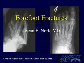
Forefoot Reconstruction. Michael Lapner 2009 04 15. Sections. Hallux Valgus Hallus Varus Hallux Rigidus Lesser Toe Deformities Intractable plantar Keratosis Freiberg Infraction Bunionette. Hallux Valgus. Etiology & Pathoanatomy. Etiology. Cause debated multifactorial

E N D
Forefoot Reconstruction • Michael Lapner • 2009 04 15
Sections • Hallux Valgus • Hallus Varus • Hallux Rigidus • Lesser Toe Deformities • Intractable plantar Keratosis • Freiberg Infraction • Bunionette
Hallux Valgus • Etiology & Pathoanatomy
Etiology • Cause • debated • multifactorial • reactive ground forces/dynamic muscular • repetitive valgus force with wt bearing
med cap attenuated sesamoid ridge worn sesamoid displaced lateral structures contract
Extrinsic Factors • Ill fitting shoes • narrow box • women higher prevalence
Intrinsic factors • genetic • hypermobility first TMT joint • increase IMA 1-2 (or increased pes planus) • pes planus • shape of first metatarsal head
Clinical Presentation • Pain • difficulty with footwear • skin irritation • ulceration infection • Occupation/sports/footwear/activity level
Physical Exam • Inspection • standing • callosities • palpation • tenderness (medial eminence / under 2nd MT head)
Physical Exam • ROM • foot/ankle • motion of first MTP • motion of first TMT/compare to other side • sagittal / transverse plane • passive ROM of 1st MTP with manual correction of hallux valgus
Physical Exam • Vascular/Neurologic exam documented • appropriate consultation as required • may consider non-operative treatment
Radiographic Exam • wt bear AP • lateral • non-wt/bearing oblique • special sesamoid view (axial forefoot)
Radiographic assessment • AP • HVA • IMA 1-2 measured • DMAA
Radiographs • HVA • normal < 15 • IMA 1-2 • normal < 9 • MPV • DMAA • normal < 10
Radiographs • Prox/Distal Phalanx angle measured • hallux valgus interphalangeus • review location sesamoids • congruency • size of medial eminence • arthritis
Non Surgical Treatment • Education • wide footwear • sketch foot size • soft shoes • bunion press to relieve pressure • padding • orthoses unlikely to be of much benefit
Surgical Rx • Indication • failed non-op • pain/limited activities/inability to wear footwear • not cosmesis • expectations discussed
Principles - HV • maintain/obtain congruent joint • mild deformity - distal osteotomy • mod/severe deformity - prox osteotomy • arthrodesis/combined osteotomies
HV Mild Rx • Mild deformity • distal chevron osteotomy • Mod/severe with incongruent joint • distal soft-tissue procedure + prox osteotomy • Severe • arthrodesis • Keller (resection arthroplasty) - low function
Distal Correction • Modified McBride • medial capsulotomy with imbrication • excision of medial prominence • release of contracted deforming structures • adductor/lat capsule/transverse MT ligament
Distal Correction • distal chevron • V-shape osteotomy of MT head • capital fragment 4-6 mm translate laterally • medial joint capsule plicated • required congruent joint • no increased risk of osteonecrosis w/ lat release
Distal Correction • Biplanar chevron may be added • medial close wedge
Proximal Correction • soft tissue • correct congruency • bony • osteotomy to correct MVA/IMA12
Options-proximal • Proximal crescentic • proximal chevron • scarf • lateral close wedge • medial opening wedge
Considerations • shortening • dorsiflexion • complications • recurrence • varus • non-union
Biomechanics • Proximal chevron/long oblique/crescentic • superior fixation with screws vs K-wires
Post-Op • compressive dressing • wt bear in post-op stiff shoe • until radiographic union (8 weeks)
Arthrodesis • Dorsal incision • bony cuts made • lag screw/plate may be used (bone quality) • walk boot (12 weeks)
Hallux Valgus Interphalangeus • medial close wedge base of prox phalanx • screw/wire/staple • called Akin osteotomy
Other procedures • Cotton • medial open wedge of first cuneiform • IMA12 • Reverdin • medial distal close wedge of first MT
Lesser Toe Deformities • deformities of 2nd-5th toes • trauma • inflammatory • neurologic • ill-fit shoewear • congenital
Lesser Toes • Lesser toes • balance/pressure distribution • passive & active stabilizers
Passive stabilizers • plantar aponeurosis • capsule • plantar plate • collaterals
Active stabilizers • extrinsic muscles • EDL/FDL • intrinsic muscles • FDB EDB IO L • tibial nerve extrinsic & intrinsic • peroneal extrinsic only
Anatomy • EDL & EDB extend MTP (extensor hood) • EDL central slip into middle phalanx • medial/lateral slip insert to distal phalanx
Anatomy • EDL/EDB extend MTP PIP DIP • FDL (deep FDB) inserts at distal phalanx • strong DIP flex • weaker PIP/MTP flexors • FDB bifurcation - inserts mid phalanx
Anatomy • Lumbricals/interossei • plantar to axis of motion of MTP • flex MTP • extend PIP/DIP (dorsal extensor hood)
Clinical Presentation • symptoms with closed-toe shoes • calluses/blisters • pain under MT head • position of toes observed with wt bearing • deformity of 1st ray • flexibility • radiographs obtained
Non Surgical Treatment • Deep toe box • proper fit shoes • slings • pads • taping devices • manual stretching (no evidence)
Surgery • Failure of conservative measures • pain • inability to comply with shoe wear restrictions
Surgical Treatment • various soft-tissue and bony procedures
Complications • discussed pre-op • recurrence • incomplete correction • vascular compromise • clear communication with patient • short toes/stiff joint/prolonged swelling
Mallet Toe • Flexible • percutaneous flexor tenotomy • at DIP crease • Rigid • bony correction - resection arthroplasty • DIP arthrodesis +/- FDL tenotomy • k-wire (4-6 weeks)