Download
1 / 37
380 likes | 643 Views
P lacode ectoderm and the neural crest : d evelopment and derivatives Dr. Altdorfer. http://semmelweis.hu/anatomia/. login: educatio pw: semmelweis. 3 week-old human embryo. Placode. Placodes and neural crest. Neural crest: caudal, medial, epithelio-mesenchymal transition.

E N D
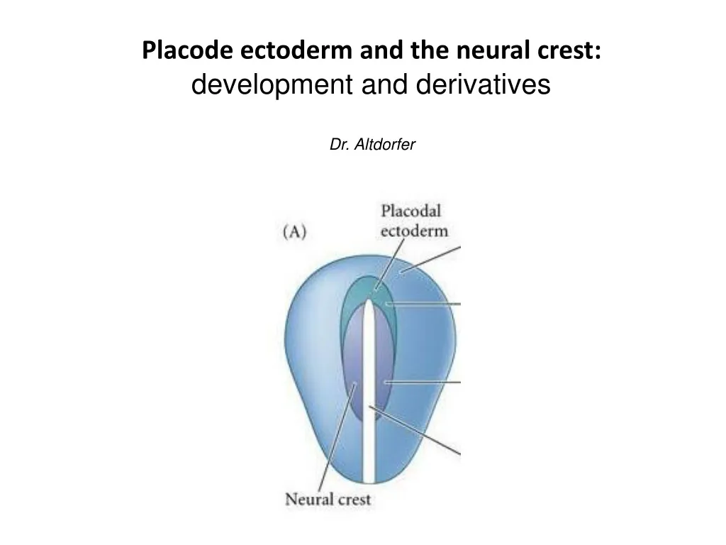
Placode ectoderm and the neural crest: development and derivatives Dr. Altdorfer
http://semmelweis.hu/anatomia/ login: educatio pw: semmelweis
Placodes and neuralcrest Neural crest: caudal, medial, epithelio-mesenchymal transition Placode: cranial, lateral, epithelialial tickenings may form vesicles Prof. Szél
Placode Derivative Hypophyseal Olfactory Lens Trigeminal Otic Epibranchial Neural plate Neural crest Ectoderm Rathke’s pouch Adenohypophysis Olfactory epithelium Lens Trigeminal ggl. (partly) Otic vesicle membranous labyrinth, spiral+vestibular ggl.* (VIII.) Epibranchial –> taste buds, geniculate ggl. (VII.), inf. ggl. of IX. and X. nerves* Brain, spinal cord ganglia, … epidermis of skin,… *Special sensory ganglia!
Neural crest cells are a multipotent progenitor population, so that the fates of specific crest populations must be controlled through environmental factors during normal development. In the trunk region the neural crest cells are divided into two groups: - Those which migrate dorsally, differentiate to melanocytes in the skin and hair follicles. - migrating through the ventral pathway can form sensory ganglia and the accompanying glial satellite cells and Schwann cells (ventrolateral migration between the dermatomyotome and sclerotome cells of the somites), or take the ventromedial pathway between sclerotome and neural tube and form sympathetic and enteric neurons, satellite cells, Schwann cells, and cells of the adrenal medulla. The neural crest cells of the head region have more opportunities: Neural derivatives: - Sensory ganglia (General sensory ones): -superior ganglia of the glossopharyngeal and vagus nerve, -part of the trigeminal ganglion (other part: from placode). • Autonomic ganglia: ciliary (III.), pterygopalatine, submandibular (VII.) and otic (IX.) ganglia (all these from 2nd rhombomer – located next to CN V. branches) “ectomesenchymal” tissue: -bones of the skull (frontal bone, parietal bone, squama of the temporal bone, nasal bone, vomer, palatine bones, maxillae and mandible) -all meninges , choroid and sclera of the eye -dentin of the teeth -connective tissue of the lacrimal, nasal, labial, palatine, oral, and salivary glands, thyroid and parathyroid glands, and thymus -connective tissue of the head (including melanocytes), cartilages, ligaments, and tendons -Tunica media of the outflow tract of the heart and the great vessels (conotruncal septum)
NC cell migration in 3 streams: • ‘trigeminal’–around 3 divisions of CN V. (1st branchial arch*+frontonasal process) • ‘hyoid’– into 2. branchial arch • ‘postotic’ (= behind otic vesicle) – into branchial arches 3-6. Ectomesenchym: bones, cartilage, conn. tissue, vessels * 2 of 3 auditory ossicles, jaw (Meckel’s cartilage)- skull… „new head” Rhombomeres 1,2,(3) Rhombomeres (3),4,(5) Rhombomeres (5),6,7,8
Neural crest cells give advantages to Vertebrates Most of the morphological and functional differences between vertebrates and other chordates occur in the head and are derived embryologically from muscularized hypomere, neural crest, and epidermal (neurogenic) placodes. In the head, the neural crest functions as mesoderm and forms connective, skeletal, and muscular tissues. Both the neural crest and the epidermal placodes form special sense organs and other neural structures. The transition to vertebrates apparently was associated with a shift from a passive to an active mode of predation, so that many of the features occurring only in vertebrates became concentrated in the head(Gans és Northcutt, Science 1983). Plasticity of neural crest cells… ecto-mesenchyme Characterictics of the vertebrate: „New Head”: Specialcomposite sense organs, complex visual organ is important in predation Jawsfor predation! NC Pigment cells against UV radiation, accomodation to environment Complex viscerocranium andchondrocraniumdevelopment (Neuron. 2003 Mar 27;37(6):895-8.A celebration of the new head and an evaluation of the new mouth.)
Derivatives of the neural crest Ganglia, peripheral nerve cells glial cells Myofibroblast, fibroblast Cartilage, bone melanocytes endocrine cells
Derivatives of the neural crest • peripheral nervous system • sympathetic, parasympathetic, sensory, enteric • Schwann cells, satellite cells • melanocytes • cartilage and bone in the head, smooth muscle, myofibroblast and fibroblast , mesectoderm • endocrine cells (adrenal medulla) • meninges • vessel wall (not endothelia) in the head region • Heart : aortico-pulmonal, conotruncal septum
Results of chimera experiments, fate mapping studies - derivatives of the neural crest enteric nervous system adrenal medulla N.M. Le Douarin / Mechanisms of Development 121 (2004) 1089–1102
Neural crest induction, current modell Signals from ectoderm: BMP, Wnt from mesoderm FGF-8 (amphibian data) high conc. of BMP: epidermal ectoderm, low: neural ectodermmedium BMP concentration defines neural crest Msx-1, Pax-3 expression starts (characteristic for NC cells) snail-1 and slug (snail-2) expression starts which is needed for EMT(epithelio-mesenchymal transition) slug is expressed at gastrulation as well! Changes in adhesion properties (adhesion molecules)
After induction: migration ECM: Fibronectin, laminin, and type IV. collagen are favorable ECM: chondroitin-sulfate: non-favorable cell-surface integrins TRUNK HEAD In the head region neural crest cells start the migration before the closure of the neural tube, not in the trunk
Neural crest divisions Circumpharyngeal pharynx heart, great vessels intestine trunk 6th somite sacral cranial
Cranial craniofacial (ecto-) mesenchyme, cartilage, bone, conn. tissue, nerve, glia Topography Cardiac arterial wall, septum aorticopulmonale Enteric vagal, sacral, parasympathetic elements Trunk melanocytes, spinal ganglia, sympathetic ganglia Prof. Szél
Neuromeres, prosomeres, rhombomeres http://www.cram.com/flashcards/neuro-47-development-of-the-nervous-system-2544719
NC cell migration in 3 streams: • ‘trigeminal’–around 3 divisions of CN V. (1st branchial arch*+frontonasal process) • ‘hyoid’– into 2. branchial arch • ‘postotic’ (= behind otic vesicle) – into branchial arches 3-6. Ectomesenchym: bones, cartilage, conn. tissue, vessels * 2 of 3 auditory ossicles, jaw (Meckel’s cartilage)- skull… „new head” Rhombomeres 1,2,(3) Rhombomeres (3),4,(5) Rhombomeres (5),6,7,8
5th and 6th prosomere level do not give rise to neural crest only 1-4 prosomeres. NC from hindbrain levels colonise 1st-3rd pharyngeal arches. Each rhombomeric and mesencephalic crest cell „remember” to the segmental code. In the pharyngeal region, the pathways of crest cell migration are closely correlated with Hoxb gene expr. Cells of the cranial crest may be patterned with level –specific instructions, whereas cells of the trunk crest are not.
DiGeorge syndrome - hoxa-3 gene defect Mesenchymal elements of cranialis crest (III-IV. pharyngeal arches) are defective Aplasia of thymus and parathyroid gland (III-IV. pharyngeal pouch), „fish-mouth” deforation (shorter philtrum), deformation of lingual and cervical muscles, hypertelorism, mal-formations of the heart Knockout mouse: short, thicke neck, lack of thymus and parathyroid gland, deformation of cardiac vessels and valves, deformation of greater horn of hyoid bone, cricoid cartilage, and epiglottis Prof. Szél
The circumpharyngeal neural crest arises in the posterior rhombencephalic region, and in the lower part of the pharynx, emigrating circumpharyngeal crest cells pass behind the sixth pharyngeal arch. Neural crest cells from the anterior rhombencephalon to the level of somite 5 emigrate from the circumpharyngeal crest as a stream, called the cardiac crest, toward the developing heart and aortic arches, whereas other neural crest cells from the levels of somites 1 to 7 constitute the vagal crest and migrate into the developing gut as precursors of the parasympathetic innervation of the digestive tract.
Results of chimera experiments, fate mapping studies - derivatives of the neural crest enteric nervous system adrenal medulla N.M. Le Douarin / Mechanisms of Development 121 (2004) 1089–1102
19-day old chicken embryo colon Remak ganglion plexus myentericus plexus submucosus Dr. Nagy
Hirschprung’s disease (megacolon congenitum aganglionare) -developmental anomaly; Affects 1:5000 human infants. • complete absence of ENS in the distal bowel (no relaxation!); proximal colon becomesdistended • 90% are diagnosed as newborns. • Failure to pass stool within 1st 2 days of life, abdomen distended, vomiting Dr. Nagy
Trunk neural crest Migratory pathways ventrolateral pathway (anterior somite: sensory ganglia) dorsolateral pathway (melanocytes) ventral pathway (sympathico-adrenal) Prof. Szél
References Schoenwolf, Bleyl, Brauer, Francis-West: Larsen’s Human Embryology, Elsevier S. F. Gilbert: Developmental Biology, Sinauer associates, Inc. Publishers T. W. Sadler: Langman’s Medical Embryology, Williams & Wilkins B. M. Carlson: Human Embyology and Developmental Biology Lectures of Anatomy Department (Prof. Szél Á., Prof. Csillag A., Prof. Kálmán M. Dr. Nagy N., Dr. Kocsis K., Dr. H.-Minkó K.) Neural regulation of human life processes – from the neuron to the behaviour. Interdisciplinary teaching material concerning the structure, function and clinical aspects of the nervous system for students of medicine, health and life sciences in Hungary University of Pecs; Dialóg Campus Publishing-Nordex Kft.