Download
1 / 25
250 likes | 403 Views
Biology of Human Aging. Chapter 4 The Integumentary System. Outline . Review of structure and Function Epidermis / Dermis 2. Age-related Changes Epidermal Changes Dermal Changes Hypodermal Changes 3. Age-Related Dysfunctions

E N D
Biology of Human Aging Chapter 4 The Integumentary System
Outline • Review of structure and Function • Epidermis / Dermis • 2. Age-related Changes • Epidermal Changes • Dermal Changes • Hypodermal Changes • 3. Age-Related Dysfunctions • Lentigo, Senile Purpura, Senile Angioma, Acrochordon, Senile Pruritus, Senile keratosis, Seborrheic Keratosis, Herpes Zoster, Decubitus Ulcers • 4. Skin Cancers • Basal Cell Carcinoma • Squamous Cell Carcinoma • Malignant Melanoma • Secondary Skin Cancer
Introduction/Background • Integumentary system comprises the skin, hair, nails & various glands located in the skin. • Age change in general appearance of skin wrinkles & sags • Coupled w/ gradual graying of hair reminder of aging changes creams & hair dyes won’t actually delay aging process • Plastic surgery removes some of the aging changes still not helpful! • Factors affecting rate and degree of changes in integumentary system: • Intrinsic factors • Heredity • Dietary habits • Levels of various hormones • extrinsic factors • Sun and wind (increase potential for skin cancer) use of protective sun-shield lotions
Other factors contributing to aging of skin: • Occupation: Extended sun exposure (photoaging) • Recreational preference: Boating or sunbathing • Exposure to sun is the major cause of aging changes in the skin
Review of Structure and Function • Single most important function: help to maintain a stable internal environment (homeostasis) normal functioning of various cells • Protective covering prevents water loss • Partial barrier blocks the entrance of microorganisms • Pigment cells protect against UV radiation; (Vitamin D) • Sweat glands and network of blood vessels regulate temperature • Temperature rise: • blood vessel dilation higher volume of blood to surface heat is lost by radiation from blood to environment • Increase in sweat gland activity skin surface becomes wet body heat loss by evaporation • Provides information on external environment to nervous system (through receptors sensitive topain, temperature, touch)
Review of Structure and Function • Process drugs and compounds (smoking patches, seasickness) this function is similar to the function of liver • Part of the immune system (hormone that enhances the growth and development of T cells residing in skin)
The Integumentary System Skin: major component of the integumentary system, separates body from external environment via an interrupted covering over entire body • Consists of three major regions: • Epidermis – outermost superficial region • Dermis – middle region • Hypodermis (superficial fascia) – deepest region (a layer of loose connective tissue; attaches dermis to underlying muscles, also fat deposition provides padding & also fat storage)
Skin (Integument) Figure 5.1
Epidermis • Consists of several layers of thin, flat cells (squamous cells) form stratified squamous epithelium • Under constant pressure or friction thickens soles of the feet & palms of the hand calluses and corns • No blood vessels or nerve fibers; nutrient and waste diffusion • Inadequate supply (gas & nutrients) to outer cells dead cells gradual replacement of cytoplasm with Keratin, • Outer-most layer of epidermis composed of thin, dead cells • All of the cells of epidermis are replaced every 28 days • Skin color: determined by amount & distribution of melanin • Dark skinned: contain more melanin • Light-skinned people: reddish hue due to blood vessels • Oriental people: variation of melanin that causes their epidermis to have yellowish hue
Cells of the Epidermis • Keratinocytes – produce the fibrous protein keratin • Melanocytes – produce the brown pigment melanin • Langerhans’ cells – epidermal macrophages that help activate the immune system • Merkel cells – function as touch receptors in association with sensory nerve endings
Functions • Waterproofing • Protection from abrasion and penetration • Rendering the body relatively insensitive to biological, chemical, and physical assaults
Dermis • Located immediately beneath the epidermis, thicker than epidermis • Second major skin region containing strong, flexible connective tissue (collagenous& elastic fibers) • Well-supplied with blood vessels, nerves, sweat glands, oil-secreting sebaceous glands • Specialized receptors provide information concerning touch, pain, pressure, temperature changes to nervous system • Cell types include fibroblasts, macrophages, and occasionally white blood cells • Composed of two layers – papillary and reticular
Layers of the Dermis: Papillary Layer • Papillary layer • Areolar connective tissue with collagen and elastic fibers • Dermal papillae contain capillary loops, Meissner’s corpuscles, and free nerve endings
Layers of the Dermis: Reticular Layer • Reticular layer • Accounts for approximately 80% of the thickness of the skin • Collagen fibers in this layer add strength and resiliency to the skin • Elastin fibers provide stretch-recoil properties
Hypodermis • Subcutaneous layer deep to the skin • Composed of adipose and areolar connective tissue
Skin Color • Three pigments contribute to skin color • Melanin – yellow to reddish-brown to black pigment, responsible for dark skin colors • Freckles and pigmented moles – result from local accumulations of melanin
Sweat Glands and Sebaceous Glands • Sweat Glands • Different types prevent overheating of the body • Sebaceous Glands • Soften skin when stimulated by hormones • Secrete an oily secretion calledsebum
Sebaceous Glands • Simple alveolar glands found all over the body • Soften skin when stimulated by hormones • Secrete an oily secretion called sebum
Functions of the Integumentary System • Protection – chemical, physical, and mechanical barrier • Body temperature regulation is accomplished by: • Dilation (cooling) and constriction (warming) of dermal vessels • Increasing sweat gland secretions to cool the body • Cutaneous sensation – exo-receptors sense touch & pain
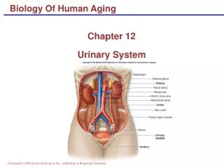
Functions of the Integumentary System • Metabolic Functions – synthesis of vitamin D in dermal blood vessels • Blood Reservoir – skin blood vessels store up to 5% of the body’s blood volume • Excretion– limited amounts of nitrogenous wastes are eliminated from the body in sweat
Developmental Aspects of the Integument: Old Age • Epidermal replacement of cells slows and skin becomes thinner • Skin becomes dry and itchy • Subcutaneous fat layer diminishes, leading to intolerance of cold • Decreased elasticity and loss of subcutaneous tissue leads to wrinkles • Decreased numbers of melanocytes and Langerhans’ cells increase the risk of skin cancer
Age-related changes • Epidermal changes • Thinner • Permeability of the surface cells increased • Larger melanocytes and grouped together; dark pigment plaques (age spots) • Decrease in the number of immune cells in skin with aging
Age-related changes • Dermal Changes • Number of fibroblast and fibers is reduced • Thin and somewhat translucent • Elastic fibers become less resilient • Slight calcification & formation of cross-links • Reduce in numbers and gradual atrophy of sweat & sebaceous gland • temp. regulation problem slow in growth of fingernails • General loss of body hair • Reduction of pigment in the hair with aging (heredity factors) • Changes in dermal sensory receptor (pain, temp, touch, etc)
Age-related changes • Hypodermal changes (subcutaneous tissue) • General loss of fat (most obvious in face and limbs) • Cause of wrinkles • Loss of padding reduction in blood supply to the skin pressure sores when bedridden • Temperature regulation (older individuals feel chilly most of the time) • Modern Maturity Magazine • “old age is when, upon getting out of the bathtub, you notice that the full-length mirror is steamed up • – and you are glad of it”
Age-related Dysfunctions Lentigo: after 50, dark-brown irregular areas, increased melanin, no tendency to malignancy Senile Purpura: irregularly shaped purple patches, forearm and back of hands, minimal Senile Angiomas: 75% over 70, elevated clusters of dilated capillaries, red spot, Acrochordon: small pendulous skin growth (cutaneous tags),chest, neck, eyelids Senile pruritus:loss of water, oil-secreting sebaceous glands, sweat glands, cracks(itching) Senile keratosis:(actinic keratosis) localized red areas of skin, soft-thicken-scaly-yellow brown precancerous Seborrheic keratosis:formation of benign epidermal tumors, no precancerous, face, chest, back Herpes Zoster:viral disease, shingles, same virus that causes small-pox, attacks sensory nerve Decubitus Ulcers:pressure sores cavities of dead tissue form in skin, bedridden or immobilized Skin cancer: malignant vs. benign, metastasize, early detection, melanoma vs. non-melanoma Basal cell carcinoma:most common, from cells in deepest or basal layer of epidermis Squamous cell carcinoma: develops from squamous EP, less common than basal, malignant Malignant melanoma: highly malignant, potentially dangerous, usually develops in melanins Secondary skin cancer:originate in other areas of the body