Download

1 / 59
650 likes | 1.14k Views
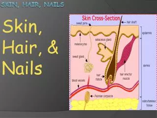
NAILS. Anatomy. Nail plate: is the hard and translucent portion, composed of keratin Its free edge is the part that extends past the finger 2. Lunula : is the crescent shaped whitish area of the nail bed

E N D
Anatomy • Nail plate: is the hard and translucent portion, composed of keratin Its free edge is the part that extends past the finger 2. Lunula : is the crescent shaped whitish area of the nail bed 3. Eponychium or cuticle, is the fold of skin at the proximal end of the nail
4. Nail fold : a fold of hard skin overlapping the base and sides of the nail 5. Nail matrix : situated directly below the cuticle. New cells form here to produce the nail plate. It also contains blood vessels and nerves. If the matrix is damaged the nail will grow deformed 6. Nail bed : It is continuation of the matrix and the part that the nail plate rests on
7. Hyponychium: is the attach- ment between the skin of the finger or toe and the distal end of the nail
Nail growth and factors affecting it • The nail can take between 5 - 6 months to grow from the matrix to the free edge • In children the growth is rapid 6 – 8 weeks • In adults nails grow approximately by 1 mm a week • Nail growth is quicker: · During pregnancy · In the summer than in the winter · On the hands than the feet • If the matrix is damaged by trauma, the nail will grow back deformed
Nail growth is inversely proportional to age • With advancing years the nail plate becomes paler and opaque and white nails similar to those seen in cirrhosis, uraemia and hypoalbuminaemia may be seen in normal old persons • Longitudinal ridging is present to some degree in most people after 50 years of age
I-Nail disorders 1.Congenital 2.Traumatic 3.Infectious 4.Neoplastic
1.Congenital Anonychia • Some or all the fingernails and toenails are absent without significant bone anomalies • It is a rare condition and may have an autosomal dominant inheritance pattern
Nail patella syndrome • A rare autosomal dominant syndrome • There is a tetrad of fingernail dysplasia with triangular lunula, absent or hypoplastic patellae, posterior iliac horns, deformation of the radial heads
Pachyonychia congenita • A rare autosomal dominant disease • There is hypertrophy of the nails associated in some cases with nail bed and hyponychia hyperkeratosis • May be associated with palmoplanter hyperkeratosis, warty skin lesions on the limbs, hyperhidrosis and lustreless and kinky scalp hair
2.Traumatic Acute trauma • It is classified with respect to severity, ranging from a small hematoma to digit amputation
Chronic repetitive trauma Nail biting • It is found in 60% of children, 45% of adolescents and 10% of adults • The majority of moderate nail biters have no associated psychiatric disorder • Patients are susceptible to bacterial and viral infections producing whitlows. It may also result in the transportation of bacteria that are buried under the surface of the nail, or pinworms from anus region to mouth • Regarding social effects the aesthetic aspect of the nail may affect employability, self-esteem, and interaction with other people
The nails are typically short, with up to 50% of thenail bed exposed. The free edge may be even or ragged. Surface change may include splitting of the nail into layers or a sand-papered effect, and the nail may acquire a brown longitudinal streak
Cure relies largely on the motivation of the patient • Local antiseptics and antimicrobial ointments may help settle the infection secondary to nail-unit damage • Antidepressants and behavioural therapy have been used with some success in limited studies
Hangnail • Trim the skin of the hangnail with a pair of clean scissors. • These are due to hard pieces of epidermis breaking away from the lateral nail folds • Although often due to nail biting, they may result from many other minor injuries • The splits may be painful when they penetrate to the underlying dermis • They should be removed with sharp, pointed scissors
Trauma by footwear • Due to repetitive trauma by footwear,the great toenail becomes thickened, yellow and twisted (onycho- gryphosis). It is most commonly seen in the elderly
Treatment is either : - Radical by surgical removal of the nail and matrix and is recommended in young persons with good circulation - Palliative treatment requires regular paring and trimming of the affected nails, usually by a chiropodist
Ingrowing toenail • The lateral nail fold of the great toe is penetrated by the edge of the nail plate, resulting in pain, sepsis and, later, the formation of granulation tissue
The main cause for the deformity is compression of the toe from the side due to ill-fitting footwear • In infancy, ingrowing toenail most commonly occurs before shoes are worn, associated with crawling, or wearing undersized jumpsuits • Treatment is by: -Avoiding tight-fitting or high-heeled shoes - oral antibiotics or topical antibiotic ointments combined with local anaesthetic agents help to heal the toe faster and also provide pain relief - Surgical removal of the ingrown toenail may be required if the condition worsens
3-Nail infections • Paronychia • It is a soft tissue infection around a fingernail • It results from a breakdown of the protective barrier between the nail and the nail fold leading to entry of bacteria or fungi into the area • It is the most common hand infection
Etiology Acute paronychia • Usually results from a minor(e.g. nail biting) trauma that breaks down the physical barrier between the nail bed and the nail allowing the infiltration of infectious organisms • S. aureus is the most common infecting organism. Organisms, such as Streptococcus and Pseudomonas species, gram-negative bacteria, and anaerobic bacteria are other causative organisms
Chronic paronychia • It is primarily caused by a mixture of C.albicans and bacteria • It can also be a complication of eczema • Most often it occurs in persons whose hands are repeatedly exposed to moist environments or in those who have prolonged and repeated contact with irritants such as mild acids, mild alkalis, or other chemicals. People who are most susceptible include housekeepers, dishwashers, and swimmers
Clinically Acute paronychia • The presenting complaints are pain, tenderness, and swelling in one of the lateral folds of the nail • The affected area often appears erythematous and swollen • In more advanced cases, pus may collect under the skin of the lateral fold
If untreated, the infection can extend into the eponychium, both lateral folds and in severe cases, the infection may track proximally under the skin of the finger and volarly to produce a felon (whitlow) which is an abscess involving the bulbous distal end of a finger
Chronic paronychia • Patients complain of symptoms lasting 6 weeks or longer • The nail folds become swollen, erythematous, and tender without fluctuance • Eventually, the nail plates become thickened and discolored, with pronounced transverse ridges • The cuticles and nail folds may separate from the nail plate, forming a space for the invasion of various microorganisms
Treatment Acute paronychia • Oral antibiotics with gram-positive coverage against S aureus, such as amoxicillin and clavulanic acid (Augmentin) or clindamycin (Dalacine C), are usually administered concomitantly with warm water soaks 3-4 times/day • Dalacine C and Augmentin also have anaerobic activity; therefore, they are useful in treating patients with paronychia due to oral anaerobes contracted through nail biting
If the paronychia does not resolve or if it progresses to an abscess, it should be drained promptly
Chronic paronychia • The avoidance of moist environments or skin irritants is essential for recovery • Because of the 'mixed' etiology of the inflammation, many clinicians use antibiotic-anticandida-steroid creams combinations. Oral ketoconazole or fluconazole may be added in more severe cases • Patients with diabetes and those who are immunocompromised need more aggressive treatment because the response to therapy is slower in these patients than in others
Cryosurgery, using liquid nitrogen spray to the nail folds, or surgical removal of the proximal nail fold and adjacent part of the lateral nail folds, may cure recalcitrant cases
Pseudomonas infection • It is always a complication of onycholysis or chronic paronychia • The nail plate has a characteristic bluish-black or green color due to accumulation of the pigment pyocyanin below the nail which may remain after the organism has been removed • Treatment is as described for paronychia • Gentamicin or sulphacetamide eye drops can be used to eradicate the colonization in resistant cases
4-Tumors • Warts • Fibrokeratoma:They arise in the periunguium and have a hyper- keratotic tip and narrow base • Subungual exostosis: It is a benign bony outgrowth of the distal part of the toe
There is pain, which may be spontaneous or evoked by mild trauma or temperature change. Nail-plate changes depend on the location of the tumour. Matrix tumours cause splitting and distortion of the nail plate. Nail bed lesions are most likely to appear asbluish or red foci of 1-5mm diameter beneath the nail Glomus tumour:
Squamous cell carcinoma: There are hype-rkeratotic, warty changes, erosions and fissuring, macerated cuticle, periungual swelling, erythema • Melanocytic nevi: Present as longitudinal melanonychia
Malignant melanoma:There are many features that should suggest the possibility of malignant melanoma: • The presence of brown-black periungual pigmentation • The pigmentation develops in a single digit in adult life
The pigmentation is evolving to become darker and broader and has blurred edges • Longitudinal melanonychia(75% of cases)
Dermatoses affecting the nails 1-Psoriasis • Psoriasis is the most common disorder affecting fingernails (50% of psoriatics) • Pitting:Punctate surface depressions • Onycholysis:Separation of the nail from the nail bed either proximally or distally
Subungual hyperkeratosis: most marked distally and extends proximally • Splinter hemorrhage:may be due to the increased capillary prominence and in nail-bed dermis
2-Darier's disease • There are white and red longitudinal lines and distal notching
3-Lichen planus • Nail involvement occurs in 10% of individuals with disseminated LP • Nail apparatus involvement may be the only manifestation • One, several, or all 20 nails may be involved ("twenty-nail syndrome," where there is loss of all 20 nails without any other evidence of lichen planus elsewhere on the body)
There is thinning of the nail plate and longitudinal ridging • Typically, the area of the lunula is more elevated than the more distal portion
4-Alopecia areata • The nail plate is rough with a "hammered brass" appearance
5-Eczema • Severe pompholyx around the nail folds may cause nail dystrophy, resulting in irregular ridges
Nail signs in systemic disease I-Abnormalities of shape 1-Clubbing • It is the bulbous uniform swelling of the soft tissue of the terminal phalanx of a digit with subsequent loss of the normal angle between the nail and the nail bed • It is due to vasodilation of the digit blood vessels, the cause of which is unknown
Causes : 1-Primary (idiopathic)clubbing e.g. familial clubbing 2-Secondary clubbing include the following: • Pulmonary disease e.g. Lung cancer, cystic fibrosis • Cardiac disease e.g. Cyanotic congenital heart disease • GIT disease e.g. inflammatory bowel disease • Skin disease e.g. Pachydermoperiostosis • Malignancies e.g. Thyroid cancer, Hodgkin disease, leukemia • Miscellaneous conditions e.g. Acromegaly, pregnancy, and hypoxemia possibly related to long-term smoking of cannabis
2-Koilonychia • Nails become concave (spoon-shaped) • It is common in infancy as a benign feature of the great toenail • The most common systemic association is with iron deficiency
II-Changes in nail surface Beau's lines • They are deep grooved lines that run from side to side on the fingernail due to a tempo- rary cessation of cell division in the nail matrix • This may be caused by an infection or trauma in the nail matrix
Systemic reasons include: coronary occlusion, hypocalcaemia, diabetes, certain drugs - including beta blockers
Muehrcke's lines: • They are superficial (not grooved as beau’s line) white lines that extend all the way across the nail and lie parallel to the lunula • The lines are actually in the vascular nail bed underneath the nail plate, and so they do not move with nail growth and disappear when pressure is placed over the nail
The appearance of Meuhrcke's lines is nonspecific, but they are often associated with decreased protein synthesis, which may occur during periods of metabolic stress (e.g., after chemotherapy) and in hypoalbuminemic states such as the nephrotic syndrome
'True' Leukonychia • It is the most common form of leukonychia, small white spots affecting one or two nails • Picking and biting of the nails are a prominent cause in young children and nail biters • In most cases, they disappear after around eight months, which is the amount of time necessary for nails to regrow completely