Download
1 / 138
1.41k likes | 1.64k Views
CARDIOVASCULAR SYSTEM. Dr.Vindya Rajakaruna MBBS (COLOMBO). CVS consists of:. Heart Blood vessels. heart arteries arterioles veinsvenules capillaries. What is the function of CVS. Circulate blood throughout entire body for Transport of oxygen to cells

E N D
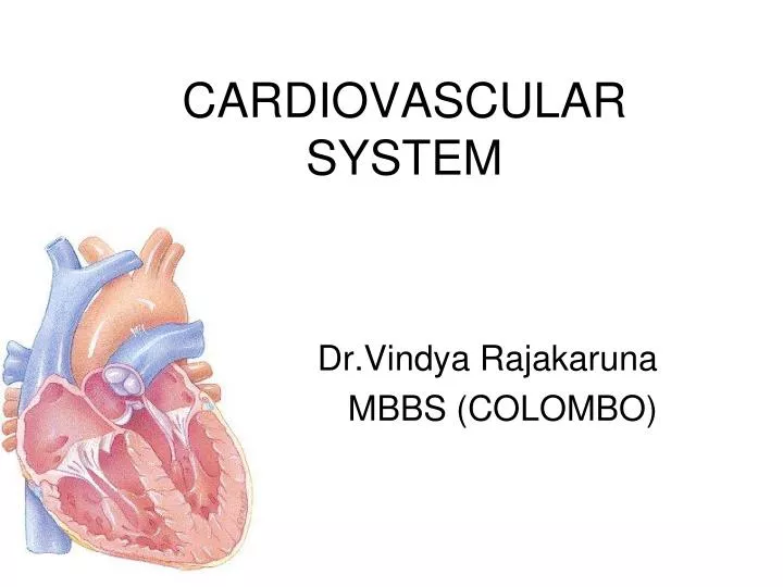
CARDIOVASCULAR SYSTEM Dr.Vindya Rajakaruna MBBS (COLOMBO)
CVS consists of: • Heart • Blood vessels heartarteries arterioles veinsvenules capillaries
What is the function of CVS • Circulate blood throughout entire body for • Transport of oxygen to cells • Transport of CO2 away from cells • Transport of nutrients (glucose) to cells • Movement of immune system components (cells, antibodies) • Transport of endocrine gland secretions
Heart • The heart is a cone-shaped, muscular organ • An adult human heart weighs between 200 and 425 grams (7 and 15 ounces) and is slightly larger than a fist. • The heart is located between the lungs in the middle of the chest, behind and slightly to the left of the sternum and in front of the spine
A double-layered membrane called the pericardium surrounds the heart like a sac. Layers of the heart wall • Three layers of tissue form the heart wall. The outer layer of the heart wall is the epicardium, the middle layer is the myocardium, and the inner layer is the endocardium.
Epicardium • The epicardium is the outer layer of the wall of the heart. It is composed of connective tissue ( mainly Adipose Tissue) covered by epithelium. • Coronary vessels and cardiac nerves • The epicardium is also known as the visceral pericardium • Provides an outer protective layer for the heart.
Cardiac Muscle/Myocardium Intercalated Disc
Myocardium is the muscular middle layer of the wall of the heart. It is composed of spontaneously contracting cardiac muscle fibers which allow the heart to contract. • Stimulates heart contractions to pump blood from the ventricles and relaxes the heart to allow the artria to receive blood.
Endocardium The endocardium is the inner layer of the heart. It consists of epithelial tissue and connective tissue. • Lines the inner cavities of the heart, covers heart valves and is continuous with the inner lining of blood vessels. • Purkinje fibers are located in the endocardium. They participate in the contraction of the heart muscle.
Pericardium • The pericardium is the fluid filled sac that surrounds the heart and the proximal ends of the aorta, vena cava, and the pulmonary artery. • Pericardial Membranes • The pericardium is divided into two layers:Fibrous Pericardium - the outer fibrous sac that covers the heart. Serous Pericaridum – The membranous covering on the outside
The serous pericardium is divided in to • Parietal Pericardium • Visceral Pericardium
Functions of Pericardium • Keeps the heart contained in the chest cavity. • Prevents the heart from over expanding when blood volume increases. • Limits heart motion.
Chambers of the heart • The human heart has four chambers. The upper chambers are called the left and right atria, and the lower chambers are called the left and right ventricles. • A wall of muscle called the septum separates the left and right atria and the left and right ventricles.
The two atria are thin-walled chambers that receive blood from the veins: the right atrium receives deoxygenated blood from systemic veins, while the left atrium receives oxygenated blood from the pulmonary veins. • The two ventricles are thick-walled chambers that forcefully pump blood out of the heart.
Differences in thickness of the heart chamber walls are due to variations in the amount of myocardium present, which reflects the amount of force each chamber is required to generate. • The left ventricle is the largest and strongest chamber
Valves • Valves are flap-like structures that allow blood to flow in one direction. The heart has two kinds of valves, atrioventricular and semilunar valves.
Atrioventricular Valves • The atrioventricular valves are thin structures that are composed of endocardium and connective tissue. They are located between the atria and the ventricles. Mitral Valve Tricuspid Valve
Semilunar Valves • The semilunar valves are flaps of endocardium and connective tissue reinforced by fibers which prevent the valves from turning inside out. • They are shaped like a half moon, hence the name semilunar (semi-, -lunar).
The semilunar valves are located between the aorta and the left ventricle and between the pulmonary artery and the right ventricle. Aortic Valve Pulmonary Valve
Function of the Heart Right Side of the Heart • The right side of the heart receives de-oxygenated blood from the body tissues (from the upper- and lower-body via the Superior Vena Cava and the Inferior Vena Cava, respectively) into the right atrium.
This de-oxygenated blood passes through the tricuspid valve into the right ventricle. • This blood is then pumped under higher pressure from the right ventricle to the lungs via the pulmonary artery
Left-Hand Side of the Heart • The left-hand side of the heart receives oxygenated blood from the lungs (via the pulmonary veins) into the left atrium. • This oxygenated blood then passes through the bicuspid valve into the left ventricle.
It is then pumped to the aorta under greater pressure • This higher pressure ensures that the oxygenated blood leaving the heart via the aorta is effectively delivered to other parts of the body via the vascular system of blood vessels (incl. arteries, arterioles, and capillaries).
The Vessels Functions: • Distribution of blood • Exchange of materials with tissues • Return of blood to the heart Structure: • Most have the same basic structure: – 3 layers surrounding a hollow lumen
Arteries and veins are composed of three tunics: • tunica interna • tunica media • tunica externa • Capillaries are composed of endothelium.
Tunica Intima • innermost smooth layer • simple squamous epithelium • continuous with the endocardium • present in all vessels
Tunica Media • layer of smooth muscle - circular arrangement – contains elastin • supplied by sympathetic division of the ANS • depending on body’s needs – lumen is narrowed (vasoconstriction) or widened (vasodilation)
Tunica Externa (Adventitia) • thin layer of Connective Tissue • elastic & collagen fibres
Types of Vessels: • Arteries – carry blood away from the heart • Veins – carry blood towards the heart • Capillaries – the most important part of the vascular system; site of exchange of materials
Elastic Arteries: • Thick-walled arteries near the heart; the aorta and its major branches. • Large lumen allows low-resistance conduction of blood. • Contain lots of elastin in all three tunics. • walls stretch and recoil to propel blood • Withstand and regulate large blood pressure fluctuations.
Muscular (distributing) arteries • medium sized vessels • tunica media more smooth muscle; less elastin • major area of vaso-constriction & dilation to regulate blood flow
Arterioles (diameter of 0.3 mm or less) - smallest arteries; lead to capillary beds. - close to capillaries - single layer of muscle spiralling around the endothelial lining - regulates blood flow to capillary
Capillaries • Smallest vessels – diameter just large enough for a red blood cell • walls consist of tunica intima only (i.e. layer of endothelium) • thinness facilitates exchange of materials
Venules • Are formed when capillary beds unite. • They merge to create veins. Veins: • Thin tunica media and a thick tunica externa consisting of collagen fibers and elastic networks. • Capacitance vessels (blood reservoirs) that contain 65% of the blood supply.
Pulmonary Circulation • Deoxygenated (used) blood is pumped out of the right ventricle. • It travels through the pulmonary valve into the pulmonary artery, leaving the heart. • Deoxygenated (used) blood reaches the lungs. Here, carbon dioxide is removed from the blood and oxygen is added to it. • The fresh blood leaves the lungs.
Fresh blood enters the left atrium through the pulmonary vein. • Then, it is pumped through the mitral valve into the left ventricle. • This ends the pulmonary circulation.
Systemic Circulation • Oxygenated (fresh) blood is pumped out of the left ventricle. • It travels through the aortic valve into the aorta, leaving the heart. • Oxygenated (fresh) blood reaches the head and the body (gut, kidney, muscles), where the oxygen in it is used and replaced by carbon dioxide.
Deoxygenated blood is collected through the vena cava into the right atrium and is pumped through the tricuspid valve into the right ventricle. • This ends the systemic circulation.
Diffusion • The exchange of molecules between cells and blood occurs at the capillary level. • Capillaries are very small blood vessels with very thin walls. • Oxygen and nutrients diffuse from the blood into the cell and carbon dioxide and waste diffuse from the cell into the blood.
Cardiac Cycle • The cardiac cycles refers to the repeating pattern of contraction and relaxation of the heart • The phase of contraction is called systole and the phase of relaxation is called diastole
The right and left atria contract almost simultaneously , followed by contraction of the right and left ventricles 0.1 to 0.2 seconds later • During the time when both the atria and ventricles are relaxed, the venous return of blood fills the atria • The build up of pressure that results causes the AV valves to open and blood flow from atria to ventricles
The ventricles are about 80% filled with blood even before the atria contract • Contraction of atria adds the final 20% to the end diastolic volume , which is the total volume of blood in the ventricles at the end of diastole • Contraction of the ventricles in systole ejects about 2/3 of the blood they contain ( Stroke volume ) leaving 1/3 of blood in the ventricles as end systolic volume.
Pressure changes during cardiac cycle • When the heart is in diastole, pressure in the systemic arteries is about 80mmHg • During systole pressure in the systemic arteries is about 120mmHg.