Download
1 / 20
220 likes | 593 Views
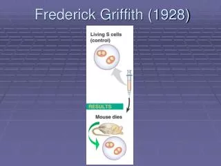
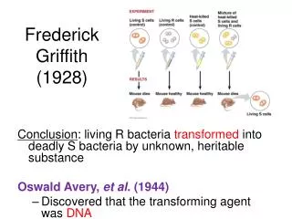
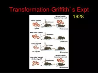
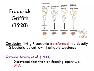
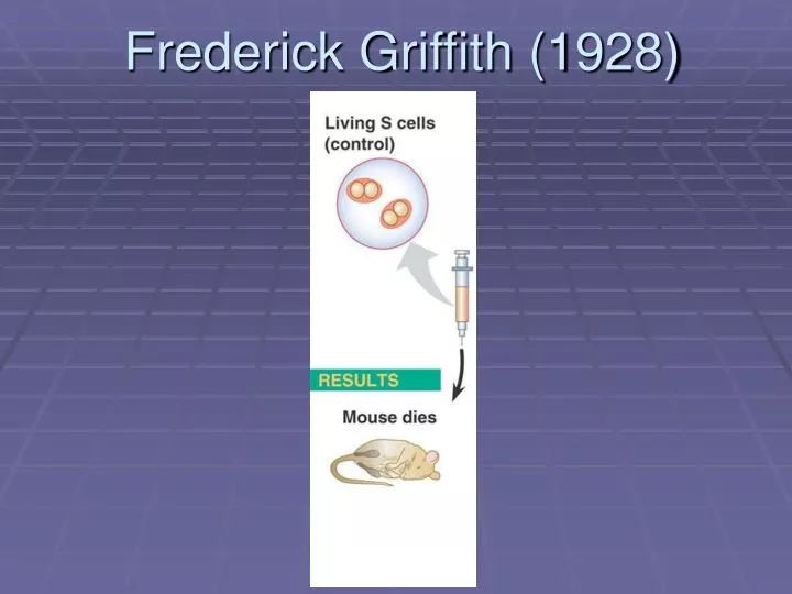
Frederick Griffith (1928). Frederick Griffith (1928). Frederick Griffith (1928). Frederick Griffith (1928). Avery, McCarty & MacLeod (1944). Only DNA (not protein, lipid or carbohydrates) TRANSFORM bacteria. Hershey & Chase (1952). “Bacteria eaters”- Phage virus particles

E N D
Avery, McCarty & MacLeod (1944) Only DNA (not protein, lipid or carbohydrates) TRANSFORM bacteria
Hershey & Chase (1952) “Bacteria eaters”- Phage virus particles (image from the 1990s)
Hershey & Chase (1952) Viruses are made of protein and nucleic acid ONLY “Something” in the virus enters the cell and gets incorporated into the genetic material (whatever that might be) of the bacteria That “something” transforms bacteria to turn them into virus production factories Protein is full of a lot of the atom Sulfur; nucleic acids have very little DNA is full of the atom phosphorus; proteins have very little
Hershey & Chase (1952) DNA “Bacteria eaters”- Phage virus particles (image from the 1990s)
Generally accepted: Nucleic acids are made of four components: Phosphate group: Sugar (deoxyribose & ribose): Nitrogenous bases: Pyrimidines (1-ring): Purines (2-ring):
Erwin Chargaff (1947) All living cells have DNA (plus many that are not living) Between species there is wide variation in the overall percentages of A, T, G & C However, in all species A = T, C = G E.g.- Human A- 30.3%, T= 30.3 %, G= 19.5%, C= 19.9% “Chargaff’s Rules” Also, a cell before meiosis has twice as much DNA as a cell (sperm, egg) after
Franklin (1951- 52) “X-ray diffraction crystallography”
Franklin (1951- 52) Image #51: Helix Repeating units “unit cell” of specific parameters Phosphates on the outside (bases inside)= “backbone” C2 form Parallel width along length
Watson & Crick (1952) Parallel width along length Backbone must be parallel Bases fill in the space between the “rungs” But how?
Watson & Crick (1952) Many “Ah-Hahs” A bonds to T (2 bonds), C bonds to G (3 bonds) and NOT vice versa Consistent with whose previous research?
“Nucleotide” Watson & Crick (1952) Many “Ah-Hahs” C2 form? All the data fit with Franklin’s, Chargaff’s, etc… conclusions
Watson & Crick (1952) All the data fit with Franklin’s, Chargaff’s, etc… conclusions And it looks good too!
Watson & Crick (1952) Part I- the model (what it looks like) Part II- how it works (the “secret of life”) “Complimentarity” & replication “The Central Dogma” & information storage
Watson & Crick (1952) DNA RNA PROTEIN “The Central Dogma” & information storage Cell Structure Enzymes Lipids Nucleic Acids Carbohydrates
Watson & Crick (1952) “Complimentarity” & replication Their Model (paper #2) “Semi-conservative” replication: