Download
1 / 92
920 likes | 926 Views
This article explores the unique characteristics of muscle tissue, skeletal muscles, nerves, and tendons, as well as the functions of bones and cartilage in the body. Learn about the naming of muscles and the different actions they perform. Discover the types of muscles and their roles in movement. Gain a better understanding of bone structure and its functions.

E N D
4 Unique Characteristics of Muscle Tissue • Excitability is equated with responsiveness. • Contractility causes the fiber to shorten resulting in either a pull on bones or the movement of specific body parts. • Elasticity is the muscle’s ability to return to its original length when tension is released. • Extensibility is capability of extending in length in response to the contraction of opposing muscle fibers.
Skeletal Muscles Skeletal muscle Vessels Nerves Tendon Epimysium Perimysium Muscle fascicle Endomysium Muscle fiber
Skeletal MusclesVoluntary muscles Under varying circumstances the degree of mobility of the attachments may be reversed; therefore, the terms origin and insertion are interchangeable
Nerve Supply of Skeletal Muscle • The nerve trunk to a muscle is a mixed nerve • about 60% is motor and 40% is sensory • it also contains some sympathetic autonomic fibers. • Motor Point • The place of entrance of the nerve into the muscle • at about the midpoint on its deep surface, often near the margin • This arrangement allows the muscle to move with minimum interference with the nerve trunk Motor neuron Motor point
Skeletal Muscle Action • Prime mover: • The chief muscle or member of a chief group of muscles responsible for a particular movement. • Antagonist: • Any muscle that opposes the action of the prime mover • Before a prime mover can contract, the antagonist muscle must be equally relaxed • this is brought about by nervous reflex inhibition Prime mover Antagonist Prime mover Antagonist Extending knee Flexion knee
Skeletal Muscle Action Prime mover • Fixator: • contracts isometrically to stabilize the origin of the prime mover so that it can act efficiently. • Synergists • assist the prime mover in performing its action. • may also assist an agonist by preventing movement at a joint and thereby stabilizing the origin of the agonist Fixator
Skeletal Muscles many muscles can act as a prime mover, an antagonist, a fixator, or a synergist, depending on the movement to be accomplished Prime mover Synergist Synergist Prime mover
Naming Muscles Skeletal muscles are named according to certain criteria A. Location- may indicate bone or body region that muscle is associated with B. Shape- Muscles often have a definitive shape, after which they are name Ex. Deltoid means triangle (and the deltoid muscle is triangular) C. Relative Size 1. Maximus= largest 2. Minimus= smallest 3. Longus= long 4. Brevis= short Ex. Gluteus maximus (larger) and minimus (smaller)
Naming Muscle D. Direction of Muscle Fibers - may reflect the direction of the fibers in relation to midline or other axis 1. Rectus= straight (runs parallel) 2. Transversus/oblique ( right angles)/ obliquely Ex. Rectus femoris- muscle that runs parallel with the femur E. Number of Origins 1. Biceps= two origins 2. Triceps= three origins 3. Quadriceps= four origins Ex. Biceps Brachii F. Location of origin and insertions 1. may be named according to the attachment points 2. Origin is always named first Ex. Sternocleidomastoid (dual origin on sternum and clavicle; insertion on mastoid process
Naming Muscle G. Action 1. Uses words such as flexor, extensor, or adductor Ex. Adductor longus on thigh adducts the thigh H. Sometimes several criteria are combined in a name. Ex. Extensor carpiradialislongus 1. muscle’s action (extensor) 2. joint it acts on (carpi= wrist) 3. Where it is (radialis= radius of forearm) 4. size (long relative to other wrist muscles
Skeletal Muscles Naming Of Skeletal Muscle Shape Size
Skeletal Muscles Naming Of Skeletal Muscle Number of heads or bellies Position
Skeletal Muscles Naming Of Skeletal Muscle Depth Attachments
Skeletal Muscles Naming Of Skeletal Muscle Action
Smooth Muscles • Composed of short muscle fibers that have a fusiform shape and single centrally located nucleus • Contraction is slow, resistant to fatigue, and usually sustained for an extended period of time. • Takes longer than skeletal muscle to contract and relax. • Contraction is under involuntary control. • Peristalsis: propulsion of contents
Cardiac Muscles Striated muscle fibers that branch and unite with each other • Conducting system of the heart: specialized cardiac fibers
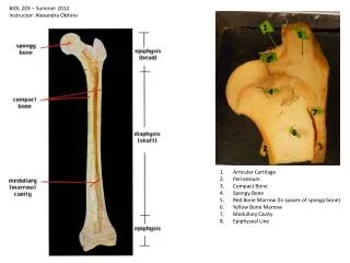
Bone • Compact bone:Solid mass • Cancellous bone: Branching network of trabecula Function of bones • The rigid supporting framework of the body • Levers for muscles • Protection to certain viscera (e.g., brain, spinal cord, heart, lung, liver, bladder) • Contain marrow, which is factory for blood cells • Storehouse of calcium and phosphate
Bone Regionally
Bone General Shape Long bones
Bone General Shape Short bones
Bone General Shape Flat bones
Bone General Shape Irregular bones
Bone General Shape Sesamoid bones
Bone • Bone marrow • Periosteum • Development of bone
BONE MARKINGS Every bump, groove, and hole has a name on your bones
Bone Markings • Two types of bone markings: • Projections (aka processes) that grow out from the bone • Depressions (cavities) that indent the bone
Joint Projections • 1) Condyle: Rounded articular projection Condyle
Joint Projections • 2) Head: bony expansion on a narrow neck • 3) Facet: smooth, nearly flat articular surface
Joint Projections • 4) Ramus: Armlike bar of bone
Ligament/Tendon Projections 1) Crest: Narrow ridge of bone (Line: smaller than a crest) 2) Epicondyle: Raised area on or above a condyle
3) Tubercle: Small rounded projection 4) Tuberosity: large rounded or roughened projection 5) Trochanter: very large, blunt projection (only on femur) Proximal Tibia
6) Spine: Sharp, pointed projection Thoracic Vertebrae
DEPRESSIONS • Allow blood vessels or nerves to pass through. 1) Meatus: (me - A- tus) Canal or tube
Depressions 2) Fossa: shallow basin 3) Fissure: narrow, slit-like opening
Depressions 4) Sinus: Cavity within a bone; filled with air and lined with mucous membranes 5) Foramen: Round or oval opening Foramen Magnum
Depressions 6) Sulcus, Groove or Furrow: a shallow depression
Projections Condyle Head Facet Ramus Crest Epicondyle Tubercle Tuberosity Trochanter Spine Depressions Meatus Fossa Fissure Sinus Sulcus or Groove or Furrow Review:
Bone Development Osteogenesis and ossification: • The process of bone tissue formation, which leads to: • The formation of the bony skeleton in embryos • Bone growth until early adulthood • Bone thickness, remodeling, and fracture repair
Bone Growth - Ossification • Cartilage template laid down. • Osteoblasts (bone building cells) located in Ossification Centers.
Bone Growth - Ossification • Primary Ossification Center in diaphasis. • Secondary Ossification Centers in epiphisis.
Bone Growth - Ossification • Grow toward one another, cartilage remains between them. • As long as cartilage remains undamaged, growth can occur.
Endochondral Ossification • Begins in the second month of development • Uses hyaline cartilage “bones” as models for bone construction • Requires breakdown of hyaline cartilage prior to ossification
Stages of Endochondral Ossification • Formation of bone collar • Cavitation of the hyaline cartilage • Invasion of internal cavities by the periosteal bud, and spongy bone formation • Formation of the medullary cavity; appearance of secondary ossification centers in the epiphyses • Ossification of the epiphyses, with hyaline cartilage remaining only in the epiphyseal plates