Download
1 / 57
590 likes | 603 Views
Ulnar-Sided Wrist Pain in the Athlete. Sarah A. Lorenzen, MOT, OTR/L Montrose M emorial H ospital. Objectives:. Identify necessary anatomy in order to properly address ulnar-sided wrist pain Identify ulnar-sided w rist injuries, causes, and symptoms

E N D
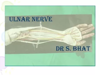
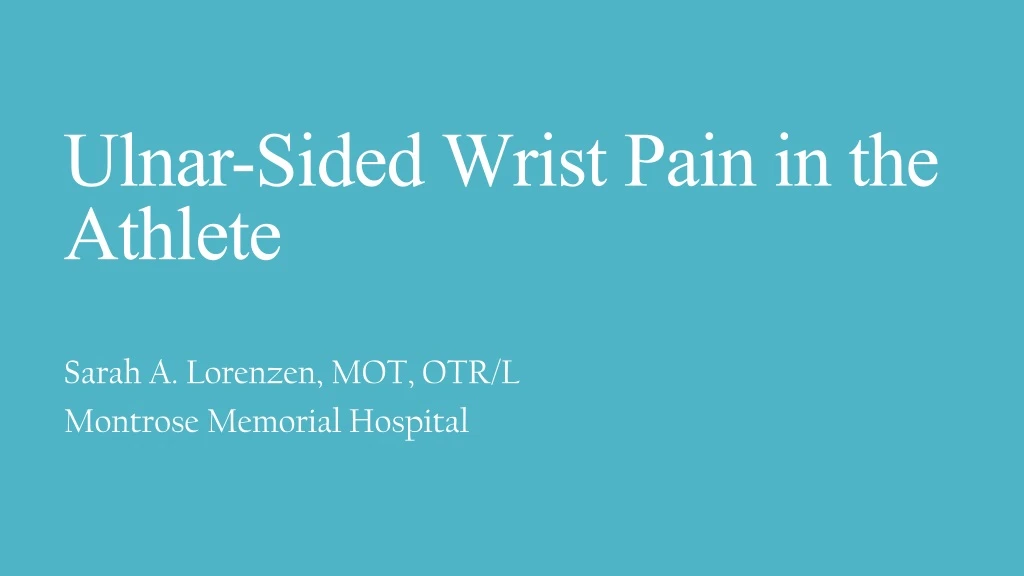
Ulnar-Sided Wrist Pain in the Athlete Sarah A. Lorenzen, MOT, OTR/L Montrose Memorial Hospital
Objectives: • Identify necessary anatomy in order to properly address ulnar-sided wrist pain • Identify ulnar-sided wrist injuries, causes, and symptoms • Address treatment considerations for ulnar-sided wrist injuries in the athlete
Anatomical Considerations: Distal Radioulnar Joint (DRUJ): • The DRUJ is formed by the sigmoid notch of the radius and the ulnar head • The axis of rotation for the forearm is from the radial head to the ulnar head. • In pronation, the radius rotates around the ulna. (Atman, 2016)
Anatomical Considerations: The Triangular Fibrocartilage Complex (TFCC): • Dorsal and Palmar Radioulnar Ligaments • Ulnocarpal Ligaments • ECU Tendon Sheath • Meniscus Homologue • Articular Disc (Triangular Fibrocartilage Proper) (Savoie, 2018)
Anatomical Considerations: The Triangular Fibrocartilage Complex (TFCC): • The TFCC is commonly known as the “Shock Absorber.” • 80% of the weightbearing load Radius • 20% of the weightbearing load Ulna
Anatomical Considerations: The DRUJ and TFCC work as a team! (Anand, 2019)
Anatomical Considerations: Anatomical Considerations: The Intrinsic Stabilizers of the DRUJ: • TFCC • Joint Capsule • Ligaments • Ulno-lunate ligament • Ulno-triquetral ligament • Distal oblique bundle (DOB) of the interosseous membrane (Bakri)
Anatomical Considerations: The Extrinsic Stabilizers of the DRUJ: • Tendon of the ECU • Sixth dorsal compartment sub-sheath • Pronator quadratus • Interosseous ligament (Wiiffles, 2012)
Ulnar-Sided Wrist Injuries: • Ulnar Variance/Abutment • Instabilities • Degeneration • Fractures • Tendinitis • Nerve Compressions
Ulnar-Sided Wrist Injuries: • Ulnar Variance: • Positive Ulnar Variance • Negative Ulnar Variance • Commonly Occurs With: • Distal radius/ulnar fractures with shortening (e.g. impaction) and angulation (FOOSH injuries) • Athletes performing power-gripping tasks associated with axial loading and rotation, such as with boxing, or gymnasts. • Sports such as boxing, mixed martial arts, and any sport that may result in a fall on an outstretched hand. • Literature suggests that 43–80% of patients following distal radius fractures, with majority of peripheral TFCC tears demonstrating DRUJ instability.
Ulnar-Sided Wrist Injuries: • What is Ulnar Variance? • Ulnar variance is the distance that the distal articular portion of the ulnar head stops proximally (negative) or extends distally (positive) compared to the articular surface of the radius.
Ulnar-Sided Wrist Injuries: Ulnar-Sided Wrist Injuries: • Positive Ulnar Variance: • Increases the stress on the ulna, lunate, and triquetrum. • A change of as little as 2mm can change the load on the carpus in pronation. • Associated conditions include: • Ulnar Impaction Syndrome/Abutment • TFCC Tears • Lunate-Triquetrum Tears Ulnar variance: Line 1 is drawn along the shaft of the radius. Two lines are drawn perpendicular to line 1. Line 2 includes the most ulnar edge of the radius. Line 3 includes the most distal edge of the ulnar seat. Ulnar variance (UV) is the distance between lines 2 and 3. (Atman, 2016)
Ulnar-Sided Wrist Injuries: • Ulnar Impaction Syndrome/Abutment: • This is a degenerative syndrome associated with positive ulnar variance. • The sequence of events includes: • Wearing of the articular disc of the TFCC • Chondromalacia of the ulnar head and ulnar aspect of the lunate • Disruption of the L-T ligament • Causes include: • Malunitedradial shortening or angulation • DRUJ ligament injuries
Ulnar-Sided Wrist Injuries: • Testing for Ulnocarpal Abutment: • GRIT Test: measures grip strength in 3 forearm positions (neutral, full supination, and pronation). The supination and pronation values are calculated as a ratio relative to neutral grip. • If the ratio is > than 1, there is a potential for impaction or the potential for an articular disc tear is high. (Ulnar Impaction Syndrome, 2019)
Ulnar-Sided Wrist Injuries: • Symptoms of Ulnar Impaction Syndrome/ Abutment: • Pain localized to the dorsal aspect of the wrist over the DRUJ or directly over the TFCC region. • Intermittent clicking sensation, activity-related swelling, decreased strength, and/or decreased motion. Proton density–weighted MRI, coronal view suggestive of ulnar impaction syndrome. Ulnar styloid process measures 8 mm (normal 2-6 mm), increased ulnar styloid index 0.61 (normal, 0.14–0.28). There is articular cartilage loss with erosion, marrow edema, subchondral cyst, and sclerosis of triquetrum and lunate. (Thomas, 2012)
Ulnar-Sided Wrist Injuries: • Negative Ulnar Variance: • This is associated with Keinbock’s Disease (avascular necrosis of the lunate). • This is of unknown etiology. Possible causes include poor vascularity and sometimes there is a history of trauma (such as a FOOSH during a sports activity). • It is predominant in 30-40 year olds. • Prevalent in football players and weight lifters.
Ulnar-Sided Wrist Injuries: • Keinbock’s Disease: • The lunate is a central bone in the wrist that is important for range of motion and strength. It works closely with the radius and ulna to help the wrist move. • Damage to the lunate can lead to pain, reduced range of motion, and sometimes arthritis. • Diagnosis can be made by reviewing the person’s history, performing a physical exam, x-rays, MRI and CT scans. • The progression of this disease is often slow, and may take quite some time to show symptoms. It may be difficult to diagnose in the early stages.
Treatment Considerations: Ulnar-Sided Wrist Injuries: • Keinbock’sDisease:
Treatment Considerations: • Ulnar Impaction Syndrome/Abutment: • A detailed history and focused physical examination are critical components for diagnosing ulnar impaction in the athlete. • The patient may experience chronic pain caused by repetitive loading of the wrist, with painful clicking or locking during pronation and supination are often present. The pain is commonly aggravated with activity, and generally relieved with rest. • ROM should be evaluated, with ulnar deviation and pronation often pain-producing. Gripping motion may elicit pain subsequent to TFCC injury. Grip strength is usually decreased compared with the unaffected wrist.Tenderness of the TFCC during palpation distal to the ulnar styloid and proximal to the pisiform, between the tendons of the ECU and flexor carpi ulnaris (FCU) indicates a positive ulnar fovea sign.
Treatment Considerations: • Ulnar Impaction Syndrome/Abutment: • Athletes may benefit from earlier intervention in an effort to return to play more rapidly • Some athletes may delay intervention depending upon their athletic aspirations and timing • During the sports season, treatment typically includes modification of activities, with NSAIDS, bracing, and therapy to decrease/control symptoms. Corticosteroid injections may be offered if extended rest is not effective. • Immobilization alone will not address altered biomechanical concerns in the athlete - ulnar variance must be adjusted with surgical treatment. • TFCC debridement and/or repair may be necessary, or arthroscopic wafer technique. Open procedures include the ulnar-shortening osteotomy (USO).
Treatment Considerations: Wrist Widget: • Ulnar Impaction Syndrome/Abutment: • Used to treat wrist pain conservatively. • Water-proof • The patient can test their weightbearing tolerance with and without it to determine if it is appropriate for their injury. • One size usually fits most wrists bilaterally!
Treatment Considerations: • Ulnar Impaction Syndrome/Abutment: • Arthroscopic debridement or repair may be appealing for midseason athletes secondary to benefit of shorter recovery time (66-87% success rate). • Following arthroscopic debridement, the patient is typically immobilized for 1-2 weeks prior to initiating ROM exercises. Strengthening begins when near normal AROM is reached. Return to modified play can occur as early as three weeks following intervention(protocol is also based on surgeon preference). • Athletes that require high-axial loading may return to sports within 4-5 weeks. • Arthroscopic wafer procedures include partial excision of the distal portion of the ulna until fluoroscopy indicates -1 to -2mm variance. The articular surfaces of the lunate and triquetrum are also evaluated, and loose cartilage may be resected. The athlete is generally splinted for 1-2 weeks and gradual return to ROM is encouraged. Patient comfort determines when they can return to full ROM. Strengthening initiated at 4 weeks. • Many patients report being symptom-free within 8 weeks, but need 6 weeks to 8 months to return to full activity.
Treatment Considerations: • Ulnar Impaction Syndrome/Abutment: Image indicates presence of ulnar styloid fracture and ulnar styloid impaction syndrome, with ulceration and cyst formation depicted within the triquetrumpre-operatively. Imaging following subsequent ulnar styloidectomy.
Ulnar-Sided Wrist Injuries: Instability: • DRUJ Instability: prominence of distal ulnar head is a sign. • “S” shape to the wrist (radial shortening, positive ulnar variance) • Other Instabilities: • LT Joint • Mid-Carpal Joint • Ulnar Carpal Joint • ECU
Ulnar-Sided Wrist Injuries: Testing for DRUJ Instability – The Piano Key Sign: • The sign is gentle downward pressure applied to the distal end of the ulna with the forearm pronated. The head moves volarly but springs back when pressure is released. • A positive sign is the “note” of pain.
Ulnar-Sided Wrist Injuries: Testing for DRUJ Instability – The Piano Key Test: • Grasp the distal ulna and move it passively in a volar and dorsal direction at extremes of pronation and supination (up to 5 mm difference may be noted). • This is done initially in neutral. • A positive test = pain, tenderness, and increased mobility relative to the uninjured side. • Also, there may be tenderness with palpation of the joint.
Ulnar-Sided Wrist Injuries: • Ulnar Compression Test: DRUJ Grind • Apply radially directed pressure on the ulnar head, into the sigmoid notch of the radius. • Combine with pronation and supination. • Compression will be painful with presence of arthritis.
Treatment Considerations: • DRUJ Instability: • Most preoperative protocols call for immobilization in a long arm Meunster brace/cast with the elbow at 90° and the forearm/wrist in neutral for six weeks. Afterwards, a thermoplastic ulnar gutter splint is used for the following six weeks. • The Modified Ulnar Gutter Forearm-based Splint is a static-progressive splint that uses progressive strapping to help stabilize the distal dorsal ulnar head into a more volar/proximal position by limiting pronation. The design of the splint positions the forearm in neutral rotation, and restricts pronation. DRUJ ligaments are able to heal by increasing contact between the radial sigmoid notch and the ulnar head, and by preventing distal ulna migration. (Monasterio, 2007)
Treatment Considerations: • DRUJ Instability: • Surgical reconstruction of the distal radioulnar ligaments, the key stabilizers of the distal radioulnarjoint, may be indicated in some cases. • After the procedure, the arm is immobilized in a long arm cast for 6 weeks with the forearm in neutral. • After the initial six weeks of long arm immobilization, the pin is removed and a custom ulnar gutter wrist splint is used for an additional 4 weeks to prevent the extremes of forearm rotation and wrist deviation. • Exercises during this time include active wrist motion, gentle hand and forearm strengthening, and active but not passive forearm rotation. Supination and pronation are regained gradually over four to six months. • Near full activity is usually permitted after four months if grip strength and wrist motion are almost recovered. • Heavy lifting and impact loading are discouraged until six months.
Treatment Considerations: • DRUJ Instability: • Direct communication with the referring physician with regard to the mechanism of injury, the affected structures, treatment to date, and any surgical intervention rendered is essential in formulating therapeutic guidelines. • Observing wound healing principles and respecting pain are basic to functional results. • Working within pain tolerance is important to avoid exacerbation of symptoms and avoid disruption of healing tissues. • Active forearm rotation exercises are initiated as soon as possible. Pain is the limiting factor in determining when A/AROM and PROM exercises may be incorporated.
Ulnar-Sided Wrist Injuries: • TFCC Lesions: • A dorsal angulation of the radius after a distal radius fracture increases the torque across the TFCC and DRUJ. • Shortening of the radius also causes significant kinematic alteration and tension on the TFCC. • Athletes who regularly rotate or put pressure on their wrists, such as tennis players or gymnasts, have a higher risk of developing a TFCC tear. • Injuries usually result from a rotational injury to the extended wrist (fall on outstretched hand).
Ulnar-Sided Wrist Injuries: • TFCC Lesions: • Common complaints include: • Decreased strength • Pain with rotation suggesting DRUJ involvement • Pain with ulnar deviation suggests TFCC pathology or ulnar impaction. Unstable central perforation of the TFCC articular disc, left wrist. (Thomas, 2012)
Ulnar-Sided Wrist Injuries: • Lesions of the Central Portion of the TFCC: • The central portion consists of chondroid fibrocartilage • The central portion of the TFCC bears the compressive forces between the ulnar head and triquetrum (which is a smooth but a mobile gliding surface). • Does not have vascularization • Lesions of the Peripheral Portion of the TFCC: • The peripheral portion is ligamentous with a thick collagen structure to bear the tensile loads (palmar and dorsal limbs). • The primary arterial supply is the dorsal branch of the anterior interosseous artery.
Ulnar-Sided Wrist Injuries: This is an image of a peripheral detachment of the TFCC from the dorsal capsule and avulsion from the radius. (Thomas, 2012)
Ulnar-Sided Wrist Injuries: • Testing for TFCC Tears: • Fovea Sign/Sulcus Sign: Palpate between the ulnar head and the triquetrum(the fovea is a groove at the ulnar styloid that serves as an attachment for the TFCC. • Sensitivity of this test: 92.5% • Specificity of this test: 86.5%
Ulnar-Sided Wrist Injuries: • Testing for TFCC Tears: • TFCC Load Test: • This test detects ulnar abutment or TFCC tears. • The test is performed when the tester ulnarly deviates and axially loads the wrist while moving in a volar and dorsal direction (or rotating the forearm). • The test is positive with pain, clicking, crepitus, and/or reproduction of symptoms. This is a graph from a study performed on Outcomes of surgically treated distal radial fractures with associated triangular fibrocartilage complex injury. The study found that disability outcomes were worse in patients with distal radial fracture where TFCC was injured. TFCC injuries are an important cofactor affecting the outcome of distal radial fractures. (Kasapinova, 2017)
Treatment Considerations: • Degeneration of the TFCC: • Degeneration of the TFCC is linked to a positive ulnar variance. • TFCC degeneration may be seen more commonly golf and tennis athletes, or people who work in jobs that require repetitive motion. • On evaluation, patients with TFCC lesions will have pain with direct palpation over the TFCC region. Symptoms may be worsened by active and resistive ulnar deviation with forearm rotation as well as forceful gripping. • X-rays and MRIs can be used to evaluate TFCC pathology. A wrist arthrography may help demonstrate tears in the TFCC. • Conservative treatment of TFCC injuries includes splinting with the forearm in neutral, NSAIDS, and activity modification.
Treatment Considerations: • Degeneration of the TFCC: • Surgical intervention can include removing up to 2/3 of the central portion of the articular disc. This will not compromise the stability of the DRUJ. • Ulnar shortening can be considered if the TFCC injury is related to positive ulnar-variance. Ulnar shortening osteotomy is a possibility. • Sutures placed through drill holes in the distal ulna, or suture anchors, can be used to treat avulsions of the ulnar attachment of the TFCC with or without an associated ulnar styloidfracture. • Other options include the Darrach procedure, the hemiresectionarthroplasty technique, and the Sauve-Kapandji procedure.
Ulnar-Sided Wrist Injuries: • ECU Tendinitis/Tenosynovitis: • Common in golfers and people who play racket sports, volleyball players, and rugby players (from the high-impact). • Evaluation of the Patient: • Patient’s history including previous sports injuries • Posture • Full ROM of the neck, shoulders, elbows, wrists, and fingers. • Evaluate for nerve compressions • Strength testing including grip, pinch, and MMT • Provacative testing as needed
Ulnar-Sided Wrist Injuries: Extensor Carpi Ulnaris Subluxation: • The ECU tendon resides in the sixth dorsal compartment of the wrist. • The tendon can subluxate or dislocate out of its groove on the dorsoulnar aspect of the ulnar head, due to issues with the ECU subsheath. • The tendon dislocates in a volar/ulnar direction with supination and ulnar deviation and relocates with pronation. • Recurrent ECU dislocation may be identified as a painful snap over the ulnar/dorsal aspect of the wrist with forearm supination. (Altman, 2016)
Treatment Considerations: • Extensor Carpi Ulnaris Tendinitis: • The patient needs to be evaluated by the therapist, which should include patient history, physical examination, and specific tests for the diagnosis. Assessment of the sport should be included, and is essential to the evaluation and treatment planning process. • Manual therapy should be included to address abnormal soft tissuemobility. • Conservative management should include immobilization of the wrist that limits ulnar deviation and supination. Traumatic subluxation of the ECU should include placing the patient in a long-arm cast with the elbow flexed to 90° and the wrist in approximately 30° of extension, radial deviation, and pronation. • Common in sports such as basketball and racquetball.
Treatment Considerations: • Extensor Carpi Ulnaris Tendinitis: • While immobilized, the patient should be instructed to avoid significant or excessive elbow flexion or supination. • Rest and splinting are the most common treatment for muscle and tendon inflammatory and overuse injuries. However, recent studies on animals indicate that activity strengthens the muscles and tendons. Inactivity weakens them by decreasing blood supply, collagen synthesis, tensile stress, and removal of metabolic enzymes. • Research indicates that rest is indicated for no longer than 7 days. • Rehab is focused on flexibility, strength, and endurance. Re-education on technique, habits while practicing the sport, and playing time are also emphasized.
Treatment Considerations: Extensor Carpi Ulnaris Tendinitis: “Experts disagree on the most efficient way to achieve improved strength and endurance after injury. They do agree that the exercise must be of sufficient intensity, duration, and frequency to develop maximal tension and stimulate muscle fatigue. This is referred to as the "overload principle" and is the basis for most progressive resistive exercise (PRE) programs.” (Lowe, 1992)
Treatment Considerations: • Hook of Hamate Fractures: • Conservative management for a nondisplaced fracture includes immobilization splinting for 6-8 weeks. • Patients should be aware that nondisplaced fractures may eventually evolve into displaced fractures. • Excision of the hook of the hamate is the common treatment for displaced fractures. • Significant splinting is usually not indicated after this surgery. ROM is usually not lost. • The patient may notice loss of grip strength, and therefore a progressive strengthening program is usually indicated. • Hypersensitivity may need to be addressed due to its close proximity to the ulnar nerve.
Treatment Considerations: Hook of Hamate Fracture in a Rock Climbing Athlete: (Lutter, 2016)
Ulnar-Sided Wrist Injuries: Guyon’s Canal Syndrome: • Guyon's canal syndrome involves injury to the distal portion of the ulnar nerve as it travels through a narrow anatomic corridor at the wrist. • Common in bicyclists. • The ulnar nerve reaches the hand via Guyon's canal to provide motor and sensory innervation to the digits. Guyon's canal is a unique location where ulnar nerve is vulnerable to compressive injury. • Etiologies include: • Ganglion Cyst • Hook of Hamate Fracture/Displacement • Tumors (e.g., lipoma) • Repetitive trauma (e.g., Cyclist's Handlebar external compression) • Aberrant Muscle (e.g., abductor digitiminimi) or excess fat tissue within the canal • Ulnar Artery Thrombosis or Aneurysm (e.g., Hypothenar Hammer Syndrome)
Ulnar-Sided Wrist Injuries: Guyon’s Canal Syndrome: The anatomic boundaries of Guyon's canal include: 1. Volar carpal ligament - the "roof" 2. Transverse carpal ligament - the “floor” 3. Pisiform, Pisohamate ligament, abductor digitiminimi - ulnar boundary 4. Hook of hamate - radial boundary
Treatment Considerations: Guyon Canal Syndrome • Evaluation should include patient’s history, including the onset of symptoms, and subjective description of numbness. Grip and pinch strength should be included, factors that make the pain better and worse, and comorbidities. • Sensory assessment should include two-point discrimination, monofilament tests, and Tinel'snerve percussion. • Positions of the upper extremity should be included, specifically wrist ROM. Wrist flexion is likely to exacerbate the symptoms. • Ulnar neuropathy is commonly known as “Cyclist’s Palsy” or “Handlebar Palsy.” In this image, the person is using a tool in prolonged wrist flexion and ulnar deviation. This can place prolonged pressure on the ulnar nerve, possibly leading to Guyon Syndrome. Modification of the tool can improve symptoms. (Johnson, 1990)
Treatment Considerations: Guyon Canal Syndrome • Conservative management of ulnar neuropathy is indicated for mild-to-moderate ulnar nerve entrapment. • Surgery is indicated when conservative management has failed. • The purpose of the surgery is to decompress the peripheral nerve – the goal is to remove the pressure to the ulnar nerve. All sites of compression must be eliminated, and the nerve should be able to glide. • Prolonged pressure on handlebars (such is in cycling) can also cause ulnar neuropathy. It is commonly termed “palsy” because the cyclist’s hand develops muscle paralysis. Treatment is recommended early-on, and should include proper fitting of the bicycle, avoiding prolonged weight-bearing through the hands, and using padded gloves.
Treatment Considerations: Guyon Canal Syndrome – Bicycle Hand Positions: Tops Drops Hoods The highest pressure over the region of interest was observed in the drops hand position. Riding in the tops hand position produce the greatest ulnar deviation. (Brown, 2014)