Download
1 / 30
300 likes | 308 Views
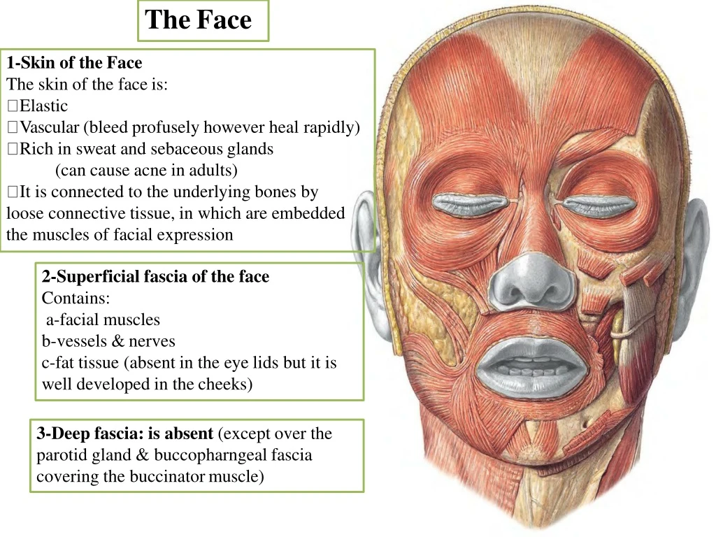
The Face. 1-Skin of the Face The skin of the face is: Elastic Vascular (bleed profusely however heal rapidly) Rich in sweat and sebaceous glands (can cause acne in adults)

E N D
TheFace • 1-Skin of theFace • The skin of the faceis: • Elastic • Vascular (bleed profusely however healrapidly) • Rich in sweat and sebaceousglands • (can cause acne inadults) • It is connected to the underlying bones by loose connective tissue, in which are embedded the muscles of facialexpression 2-Superficial fascia of theface Contains: a-facialmuscles b-vessels &nerves c-fat tissue (absent in the eye lids but it is well developed in thecheeks) 3-Deep fascia: is absent (except over the parotid gland & buccopharngeal fascia covering the buccinatormuscle)
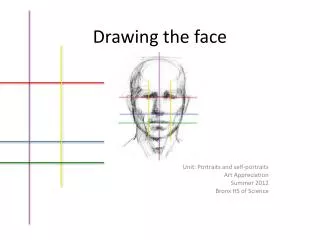
relaxed skin tensionlines Skin tension lines which follow the furrows (a line or wrinkle on a person's face) formed when the skin is relaxed are known as ‘relaxed skin tension lines’ (Borges &Alexander 1962). In the living face, these lines frequently (but not always) coincide with wrinkle lines and can therefore act as a guide in planning electiveincisions.
When lesions on the face such as scars, pigmented lesions and skincancers areexcised, the dimensions of these lesions often requireexcision as an ellipse, so that the resulting defect can be closed as astraight line. it isimportant to make the long axis of the ellipse parallel to the natural relaxed skin tension lines, so that the scar will looklike a natural skincrease To get the bestresults
Muscles of the face: muscles of the facialexpression Generalfeatures 1-They lie within the superficialfascia 2-They take their origin from the facial bones 3-They are inserted into theskin 4- They are arranged around the three openings of the face namely, the orbit, nose, and mouth either as sphincters ordilators 5- They are supplied by the facialnerve 6- Embryologically, they are originating from the mesoderm of the second branchial arch and therefore are supplied by the facialnerve 7- Can be divided into twogroups Three largemuscles Many smallmuscles
1- Three largemuscles Buccinatormuscle Orbicularis orismuscle Orbicularis occulimuscle 2-Many smallmuscles suchas: Levator labii superioris alaequenasi Levator labii superioris Zygomaticusminor Zygomaticusmajor Levator anguli oris Risorius Depressor angulioris Depressor labiiinferioris Mentalis Platysma
Buccinator Muscle of theCheek Masseter muscle, one of the muscles of mastication and its not one of the muscles of the facial expression is pierced bythe parotidduct. Nervesupply facialnerve Action: Compressesthe cheeks and lips against the teeth (prevents accumulation of foodin the vestibule of the mouth)
OrbicularisOris Nerve supply: branches of thefacial nerve Action: Compresses the lips together (closes the vestibule of the mouth)?! How you should testit?
Orbicularisoculi The orbicularis oculi is a large muscle that completely surrounds each orbital orifice and extends into eacheyelid It has two majorparts: The outer orbitalpart The inner palpebralpart Action: The orbital and palpebral parts havespecific roles to play during eyelidclosure. The palpebral part closes the eyegently whereas The orbital part closes the eye moreforcefully and produces some wrinkling on theforehead
FacialNerve As the facial nerve runs forward within the substance of the parotid salivary gland it divides into its five terminal branches 1-The temporal 2-The zygomatic 3-Thebuccal Themandibular Thecervical
Sir Charles Bell, Scottish Surgeon • First described in early 1800s based on trauma to facialnerves
Bell'spalsy Facial MuscleParalysis Damage to the facial nervein The internal acoustic meatus (bya tumor) The middle ear (by infectionor operation), The facial nerve canal (perineuritis, 4- The parotid gland (by atumor) 5- Lacerations of theface will cause distortion of the face drooping of the lower eyelid, Inability to close the eye on the affected side and the angle of the mouth will sag onthe affectedside.
Sensory Nervesof theFace The skin of theface is supplied by branchesof:the three divisions of thetrigeminal nerve except for the small area over the angle of the mandible and the parotid gland which is suppliedby the great auricular nerve (C2 and3).
OphthalmicNerve A-Frontalnerve: 1-The supratrochlearnerve supplies the skin and conjunctiva on the medial part of the upper eyelid and the skin over thelower part of the forehead, close to the medianplane. 2-The supraorbital nerve supplies the skin and conjunctiva on the central part of the upper eyelid; it also supplies the skin ofthe forehead
B-The lacrimal nerve supplies the skin and conjunctiva of the lateral part of the uppereyelid C- Nasociliarynerve The infratrochlearnerve It supplies the skin and conjunctiva on the medial part of the upper eyelid and the adjoining part of the side of the nose The external nasal nerve It supplies the skin on the side of the nose down as far as thetip
MaxillaryNerve Three branches of the nerve pass to theskin. 1-The infraorbitalnerve is a direct continuation of the maxillary nerve. It enters the orbit andappears on the face through the infraorbitalforamen. It immediately divides into numerous small branches, which radiate out from the foramen and supply the skin of the lower eyelid and cheek, the side of the nose, and the upperlip
2-The zygomaticofacialnerve passes onto the face through a small foramen on the lateral side of the zygomaticbone. It supplies the skin overthe prominence of thecheek 3-The zygomaticotemporalnerve emerges in the temporal fossa through a small foramen on the posterior surface of the zygomatic bone. It esupplies the skin over the templ
MandibularNerve The mandibular nerve supplies the skin of the lower lip, the lower part of the face, the temporal region, and part of theauricle 1-The mental nerve emerges from the mental foramen of the mandible 2-The buccalnerve 3-The auriculotemporalnerve It supplies the skin of theauricle, the external auditory meatus, the outer surface of the tympanic membrane, and the skin of the scalp above theauricle
ArterialSupply of theFace The face receives a rich blood supply from two mainvessels: 1-The facialartery 2-Superficial temporalartery
The facialartery Arises from the external carotidartery Ascends overthe submandibular salivarygland It curves around the inferior margin of the body of the mandible Passes on and in front of the anterior border of themasseter muscle(pulse)|
It runs upward in a tortuous course toward the angle of themouth It then ascends deep to the zygomaticus muscles and runs along the side of the nose to the medial angle of the eye, where it anastomoses with the terminal branches of the ophthalmic artery
2-The superficialtemporal artery ascends over the zygomatic arch, where it may be palpated just in front ofthe auricle, supplies thescalp
Supratrochlear Venous Drainage of theFace The facial vein is formed at the medial angle of the eye by the unionof The Supraorbital and Supratrochlearveins The facial vein descends behind the facial artery tothe lower margin of the body of themandible It crosses superficial to the submandibular glandand is joined by the anterior divisionof The retromandibularvein. The facial vein ends by draininginto The internal jugularvein. Importantcommunications It communicates with the pterygoidvenous plexus by the deep facialvein It communicates with the cavernous sinus bythe superior ophthalmicvein
The Supraorbitaland Supratrochlearveins Superficial temporal vein Retromandibular vein Maxillary vein Facial vein
It is connected to the superior ophthalmic vein directly through the supraorbital vein. By means of the superior ophthalmic vein, the facial vein is connectedto The cavernoussinus this connection is of a greatclinical importance because itprovides a pathway for the spread of infection from DANGEROUS AREA OF THE FACE (THE LOWER PART OF THE NOSE AND THE UPPERLIP) to the cavernoussinus Infection from the triangular area cancause Thrombosis of the cavernoussinus
Arterial Supply ofthe Scalp The arteries lie in thesuperficial fascia. A-Branches of the ophthalmic artery Thesupratrochlear Thesupraorbital B-Branch of the externalcarotid artery The superficial temporal artery The posterior auricularartery The occipitalartery
Anatomically, it is useful to remember in an emergency that all the superficial arteries supplyingthe scalp ascend from the faceand theneck. Thus, in an emergency situation, encircle the head justabove the ears and eyebrows with a tie, shoelaces, or even a piece of string and tie ittight. Then insert a pen,pencil, or stick into the loop and rotate itso that the tourniquet exerts pressure on thearteries