Download
1 / 73
760 likes | 1.04k Views
Cirrhosis of the Liver and Liver Failure. Developed by Cheryl McConnell RN MSN. Pathophysiology. Slow, insidious, progressive, chronic Fibrous bands replace normal liver structure Cell degeneration occurs Liver attempts to regenerate cells but cells are abnormal and disorganized

E N D
Cirrhosis of the Liver andLiver Failure Developed by Cheryl McConnell RN MSN
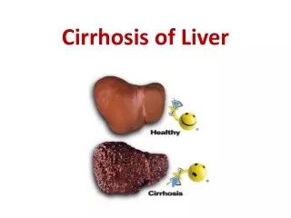
Pathophysiology • Slow, insidious, progressive, chronic • Fibrous bands replace normal liver structure • Cell degeneration occurs • Liver attempts to regenerate cells but cells are abnormal and disorganized • Causes abnormal blood and lymph flow • Results in more fibrous tissue formation
Incidence of Cirrhosis • Tenth leading cause of death in US • At least 25,000 deaths annually • Higher death rates for men than women • mortality in African Americans and Hispanics
Types of Cirrhosis • Laennec’s (alcoholic) • Postnecrotic • Biliary • Cardiac
Laennec’s Cirrhosis • Most common type of cirrhosis • Also called alcoholic or portal • Alcohol causes inflammation to liver cells • Leads to fatty deposits and hepatomegaly • Scarring formed and liver cells destroyed • Malnutrition and more alcohol accelerate the damage
Postnecrotic Cirrhosis • Caused by viral hepatitis or hepatotoxins • Scar tissue destroys liver lobes • Liver initially enlarges but then shrinks in size • 10 – 30% of all cirrhoses
Biliary Cirrhosis • Caused by chronic biliary obstruction or stasis of bile, biliary inflammation, or hepatic fibrosis • Excessive bile leads to liver cell destruction and formation of nodules in the lobes • 5 – 10% of all cirrhoses
Cardiac Cirrhosis • Seen with right sided heart failure • Liver is engorged with venous blood • Becomes enlarged, edematous, and dark • Venous congestion results in anoxia • Cell necrosis results
Diagnostic Data • AST, ALT, LDH, Alk phos • bilirubin, ammonia, • coagulation studies • Serum protein levels depend on disease • with acute liver disease • with chronic liver disease
More Diagnostics • Abdominal x-ray • Upper GI series • Angiography • Abdominal CT • EGD • Liver biopsy • Nuclear scan
Signs and Symptoms • Neurological Asterixis Paraesthesias • LOC Sensory disturbances Behavorial changes Cognitive changes • Skin Spider angiomas Palmar erythma Jaundice Pruitis hair production caput medusa pigmentation Bruising White Nails
More Signs and Symptoms • GI Abdominal pain Anorexia Ascites Diarrhea Clay colored stools Fetor hepaticus Gastritis GI bleeding N/V Varices Malnutrition
More Signs and Symptoms • Cardiovascular Dysrhythmias Portal hypertension Collateral circulation Fatigue Peripheral edema • Endocrine Gynecomastia Amenorrhea aldosterone, ADH, estrogens, glucocorticoids
More Signs and Symptoms • Respiratory Dyspnea Hypoxia • Blood Anemia DIC Thrombocytopenia WBCs Hypokalemia Hypocalcemia Hypo/Hypernatremia Hypomagnesia
More Signs and Symptoms • Immune Susceptibility to infections Leukopenia • Renal Urinary output
Complications • Portal hypertension • Ascites • Varices • Coagulation defects • Jaundice • PSE (portal systemic encephalopathy) • Hepatorenal syndrome
Portal Hypertension • Increased pressure within the portal vein • Results in obstruction of blood flow through the portal vein • Blood tries to find new ways around obstructed area = collateral circulation • Causes venous distention in entire GI tract
Ascites • Accumulation of plasma in the peritoneal cavity • Caused by increased pressure forcing fluid out of intravascular space into cavity • Plasma contains albumin so circulating proteins decreased • serum osmotic pressure • Intravascular fluid depletion stimulates kidney to conserve sodium and water = hydrostatic pressure and creates more ascites
Varices • Occur anywhere in the GI tract especially • Esophageal Hemorrhoids • Bleeding esphageal varices • Caused by thin walled veins that are irritated, distended and eventually rupture Chemical irritants Mechanical trauma • Esophagus pressure • Prone to hemorrhage – medical emergency
Coagulation Defects • Susceptible to bleeding • Bruises easily • Does not clot • Esophageal varices bleeding
Jaundice • Due to hepatocellular destruction or hepatic obstruction • Hepatocellular – cannot metabolize bilirubin so it builds up • Obstruction – clogs bile ducts so excretion is not possible
PSE • Also known as hepatic coma • Seen in end stage hepatic failure • Can be insidious or rapid onset depending on the severity of liver disease • Caused by impaired ammonia metabolism
PSE Continued • Usually protein breaks down into ammonia in GI tract, then ammonia into urea --- excreted by the kidneys • Liver cannot convert ammonia into urea • Results in serum ammonia levels • Toxic to the central nervous system