Download
1 / 1
10 likes | 126 Views
Measurement of minimum rim width (MRW) using Cirrus Optic Nerve Head Volumes in a Chinese Population. 2269 - B0073. Chen-Hsin Sun, 1 Tin A Tun, 2 John Mark S de Leon, 2 Michael JA Girard, 3, 4 Tin Aung, 2, 4 , Nicholas G Strouthidis 4, 5.

E N D
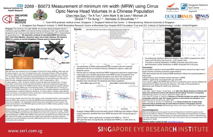
Measurement of minimum rim width (MRW) using Cirrus Optic Nerve Head Volumes in a Chinese Population 2269 - B0073 Chen-Hsin Sun,1 Tin A Tun,2 John Mark S de Leon,2 Michael JA Girard,3, 4 Tin Aung,2, 4 , Nicholas G Strouthidis 4, 5 • Duke-NUS graduate medical school, Singapore. 2. Singapore National Eye Center . 3. Bioengineering, National University of Singapore • 4. Singapore Eye Research Institute 5. NIHR Biomedical Research Centre at Moorfields Eye Hospital NHS Foundation Trust and UCL Institute of Ophthalmology, London, United Kingdom Purpose: The minimum rim width (MRW), the shortest distance between Bruch’s membrane opening (BMO) and internal limiting membrane (ILM), has recently been proposed as the most accurate Optical Coherence Tomography (OCT) measurement of neuroretinal rim.1-3 We describe a method of calculating MRW from Cirrus OCT (Carl Zeiss Meditec, Germany) optic nerve head volumes. We measure MRW in a cohort of normal Singapore Chinese eyes to characterize its distribution. Results: ILM BMO-MRW BM BMO BMO-HRW • No statistically significant difference between mean MRW computed from 36 B-scans and 4 B-scans was found (p = 0.69, paired t-test) • The plots of sectoral distribution of MRW computed using 36 B-scans demonstrated a similar distribution to MRW computed from 4 B-scans. BMO-MRW BMO-HRW • The distribution of MRW measurements was consistent with the expected distribution with the rim thickest inferiorly, followed by superior, nasal and temporal sectors • Normal eyes on average had thicker MRW compared to glaucoma suspect eyes • 2 glaucoma eyes showed significantly lower MRW across all quadrants and mean MRW beyond two standard deviations from that of the normal eyes • The superior and nasal quadrants of glaucomatous eyes showed greater deviation from normal than the temporal and inferior quadrants. • Conclusion: • It is possible to perform automated MRW measurements using Cirrus optic nerve head volumes containing automatic segmentations • We demonstrate that the sectoral MRW distribution follows the expected pattern in these eyes • Glaucomatous eyes showed marked reduction in MRW • MRW is significantly associated with age and CDR • Our results suggest that MRW derived from 4 cardinal B-scans may be sufficient for assessment for global and sectoral analyses. The utility of this parameter in glaucoma case finding will need to be assessed. Methods: 288 right eyes were selected from the Singapore Chinese Eye Study database. Mean age was 55 years (range 44 - 81) and 143 subjects were male. Intraocular Pressure (IOP) and vertical Cup to Disc Ratio (CDR) measurements (evaluated during dilated funduscopy with a 78D lens, at x 16 magnification using a slit lamp measuring graticule) were available for all eyes. Visual field (VF) tests (SITA 24-2; Humphrey Visual Field Analyzer II) were performed in 187 eyes of which 30 were unreliable (defined as fixation loss > 20% or false positive > 33% or false negative > 33%). 44 had an outside normal limits glaucoma hemifield test, 113 eyes were within normal limits and 43 eyes were borderline. 2 eyes were diagnosed as glaucoma cases (defined according to the ISGEO criteria) and 35 eyes were glaucoma suspects (defined as those participants fulfilling any of the following criteria: (1) IOP greater than 21 mm Hg, (2) VCDR > 0.6 or VCDR asymmetry > 0.2, (3) abnormal anterior segment deposits consistent with pseudoexfoliation or pigment dispersion syndrome, (4) narrow anterior chamber angle (defined in the next section), (5) peripheral anterior synechiae or other findings consistent with secondary glaucoma, and (6) known history of glaucoma). Each eye was imaged using the Cirrus OCT system using a standard 4x4x4mm cube acquisition protocol. Within each volume, the ILM and RPE/BM were automatically segmented using the native Cirrus algorithm. 36 interpolated radial B-scans were extracted from each volume. The innermost termination of the RPE/BM signal was defined as the BMO, thereby yielding 72 data points per 5 degree increment. We developed an algorithm, coded in Matlab (Mathworks, USA), which measured the shortest distance between the coordinates of each BMO point and the ILM (minimum rim width – MRW). The extracted MRW measurements were plotted across each eye quadrant to characterize the sectoral distribution of MRW. Mean MRW of each eye was calculated and associations with age, IOP, CDR and VF pattern standard deviation (PSD) were evaluated using linear regression. Furthermore sectoral distribution and mean MRW derived from the 4 'cardinal' B-scans versus 36 B-scans were compared. References: A S. Reis , Glen P. Sharpe , Hongli Yang , et al. Optic Disc Margin Anatomy in Patients with Glaucoma and Normal Controls with Spectral Domain Optical Coherence Tomography Ophthalmology, Volume 119, Issue 4, April 2012, Pages 738–747 A.S. Reis, N. O'Leary, H. Yang et al. Influence of clinically invisible, but optical coherence tomography detected, optic disc margin anatomy on neuroretinal rim evaluation Invest Ophthalmol Vis Sci, 53 (2012), pp. 1852–1860 B C. Chauhan, N. O'Leary, F A. AlMobarak, et al. Enhanced Detection of Open-angle Glaucoma with an Anatomically Accurate Optical Coherence Tomography–Derived Neuroretinal Rim Parameter Ophthalmology, Volume 120, Issue 3, March 2013, Pages 535–543 Acknowledgement: NGS acknowledges financial support from the UK Department of Health through the award made by the National Institute for Health Research to Moorfields Eye Hospital NHS Foundation Trust and UCL Institute of Ophthalmology for a Biomedical Research Centre for Ophthalmology. The views expressed in this publication are those of the authors and not necessarily those of the Department of Health • Average of Mean MRW across the entire cohort of 288 eyes was 290μm+/-60μm • Linear regression identified that mean MRW decreases with increasing age (p = 0.0016) • Vertical CDR is highly significantly correlated with MRW (p = 2X10-23) • IOP and PSD were not significantly correlated with MRW (p = 0.056 and 0.13, respectively) Primary author contact E-mail: tjensin@nus.edu.sg