Download
1 / 32
350 likes | 630 Views
Ultrasound Basics of the Hip. By Mohamed H Youssef MD Arthritis/Rehab & Pain Clinic Board certified of ABPM&R. Hip Anatomy. The hip joint is a “ball-and-socket” type joint that allows a good stability at the expense of a limited range of motion. Hip Anatomy.

E N D
Ultrasound Basics of the Hip By Mohamed H Youssef MD Arthritis/Rehab & Pain Clinic Board certified of ABPM&R
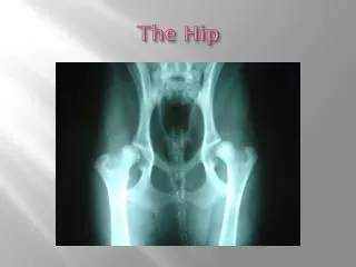
Hip Anatomy • The hip joint is a “ball-and-socket” type joint that allows a good stability at the expense of a limited range of motion.
Hip Anatomy • The hip composed of the following structures: 1-Bone structures (femoral head and acetabulum), 2-Fibrocartilaginous structures (acetabular labrum), 3-Cartilage layers covering the hip joint, 4-Capsular-ligamentous structures,
Hip Anatomy 5-Synovial joint 6-Muscles and tendons 7-Synovial bursae 8-Neurovascular structures.
Bone structures The acetabulum It is a cup-shaped cavity . The ball-shaped head of the femur fits in. The acetabularedge : circular bone flap surrounded it The lunate-surface: the most peripheral portionof the inner surface, it is used in the articulation The acetabularpit: the central portion, , accommodates the round ligament extended between the acetabulum and the femoral head surrounded by adipose tissue and vascular structures.
Bone Structures The femoral head . It is rounded in shape. • supported by the anatomical neck which is situated at an angle of about 130° to the axis of the femoral shaft in the coronal plane. • The trochanters: at the base of the neck two trochanters (the greater trochanter and the lesser trochanter) where the periarticular muscles are inserted • fovea capitisfemoris:in the center of the head there is a small depression to which the round ligament is attached.
Fibrocartilaginous structures • The acetabularlabrum: it is an axial section of the triangular fibrocartilage whose base is inserted on to the acetabular edge. • The Labrum has three main functions: 1- it inserts the capsular-ligamentous structures of the joint. 2-it increases the concavity of the acetabular fossa 3- it increases the contact area with the femoral head.
Cartilage layer covering the hip joint • The layers of articular cartilage cover the entire joint surface with the exception of the acetabular fossa and the fovea capitisfemoris. • The femoral head is covered by cartilage tissue up to the passage between the femoral head and neck.
Capsular-ligamentous structures • The articular capsule is a fibrous sheath. • Proximally it inserts on to the edge of the acetabulum and on to the acetabular labrum • distally on to the intertrochanteric line (in front) • between the third medium and distal third of the femoral neck (behind). Three peripheral thickenings of the capsule form the most important ligaments: • The iliofemoralligament •The pubofemoral ligament •The ischiofemoral ligament • The round ligament is located centrally between the acetabular fossa and the fovea capitis of the femoral head.
Synovial Joint The synovial joint lines the inner surface of the joint capsule and forms a complete sheath by surrounding the round ligament.
MUSCLES& TENDONS • 1-ANTERIOR GROUP • 2-MEDIAL GROUP • 3-LATERAL GROUP • 4-POSTERIOR GROUP
Anterior muscles • RECTUS FEMORIS • ILIOPSOAS • ILIACUS • PECTINEUS • SARTORIUS
Medial muscles • Adductor longus • Adductor brevis • Adductor magnus • Gracilis
Lateral muscles • Gluteus minimus • Gluteus medius • Gluteus maximus • Tensor fasciae latae
Posterior muscles • Semimembranosus • Semitendinosus • Long head of Biceps Femoris
Synovial bursae • Iliopsoas bursa • Peritrochantric bursa divided into: large,midium,smallbursae
What probe to use? • Curved linear probe
Where to put the probe? • 30-35 degree with the vertical • The meeting point of two imaginary lines vertical from ASIS and horizontal from ANT pubic edge. • Two fingers breadth away from femoral pulse
References • Hip: Anatomy and US technique • L. Molini,a M. Precerutti,b A. Gervasio,c F. Draghi,d and S. Bianchie, • Tom Clark ultrasound course