Download

1 / 21

E N D
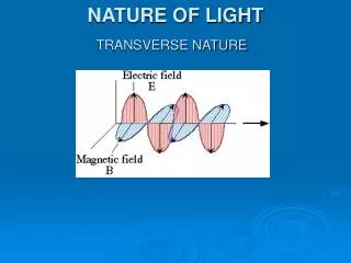
X-rays X rays are a form of electromagnetic radiation with wavelengths that range from about 10-7 to about 10-15 meter. No sharp boundary exists between X rays and ultraviolet radiation on the longer wavelength side of this range. Similarly, on the shorter wavelength side, X rays blend into that portion of the electromagnetic spectrum called gamma rays, which have even shorter wavelengths. They are invisible, are able to penetrate substantial thicknesses of matter, and can ionize matter (meaning that electrons that normally occur in an atom are stripped away from that atom). Since their discovery in 1895, X rays have become an extremely important tool in the physical and biological sciences and the fields of medicine and engineering.
X rays were discovered in 1895 by German physicist William Roentgen (1845–1923) quite by accident. Roentgen was studying the conduction of electricity through gases at low pressure when he observed that a fluorescent screen a few meters from his experiment suddenly started to glow. Roentgen concluded that the glow was caused by certain unknown rays that were given off in his experiment. Because of its unknown character, he called this radiation X rays. • Roentgen discovered that these rays were quite penetrating. They passed easily through paper, wood, and human flesh. He was actually able to insert his hand between the source and the screen and see on the screen the faint shadow of the bones in his hand. • He concluded that more dense materials such as bone absorbed more X rays than less dense material such as human flesh. He soon found that photographic plates were sensitive to X rays and was able to make the first crude X-ray photographs
Production of X rays The method by which X rays were produced in Roentgen's first experiments is basically the one still used today. As shown in the accompanying X-ray tube drawing, an X-ray tube consists of a glass tube from which air has been removed. The tube contains two electrodes, a negatively charged electrode called the cathode and a positively charged target called the anode. The two electrodes are attached to a source of direct (DC) current. When the current is turned on, electrons are ejected from the cathode. They travel through the glass tube and strike a target. The energy released when the electrons hit the target is emitted in the form of X rays. The wavelength of the X rays produced is determined by the metal used for the target and the energy of the electrons released from the cathode. X rays with higher frequencies and, therefore, higher penetrating power are known as hard X rays. Those with lower frequencies and lower penetrating power are known as soft X rays.
X-ray was the name given to the highly penetrating rays which emanated when high energy electrons struck a metal target. • Within a short time of their discovery, they were being used in medical facilities to image broken bones. We now know that they are high frequency electromagnetic rays which are produced when the electrons are suddenly decelerated - these rays are called bremsstrahlung radiation, or "braking radiation". • X-rays are also produced when electrons make transitions between lower atomic energy levels in heavy elements. X-rays produced in this way have definite energies just like other line spectra from atomic electrons. They are called characteristic x-rays since they have energies determined by the atomic energy levels.
X-rays – continuous and characteristics Characteristic x-rays are emitted from heavy elements when their electrons make transitions between the lower atomic energy levels. The characteristic x-rays emission which shown as two sharp peaks in the illustration occur when vacancies are produced in the n=1 or K-shell of the atom and electrons drop down from above to fill the gap. The x-rays produced by transitions from the n=2 to n=1 levels are called K-alpha x-rays, and those for the n=3 1 transiton are called K-beta x-rays.
Transitions to the n=2 or L-shell are designated as L x-rays (n=3 2 is L-alpha, n=4 2 is L-beta, etc. ). • The continuous distribution of x-rays which forms the base for the two sharp peaks at left is called "bremsstrahlung" radiation. • This emission spectrum is sometimes called the continuous emission spectrum because, unlike in the discrete spectrum, the energies of the photons emitted may range anywhere from zero to some maximum value. The general shape of the continuous x-ray spectrum is the same for all x-ray machines. • Characteristic x-rays are used for the investigation of crystal structure by x-ray diffraction. Crystal lattice dimensions may be determined with the use of Bragg's law in a Bragg spectrometer
Bragg’s Law • When x-rays are scattered from a crystal lattice, peaks of scattered intensity are observed which correspond to the following conditions:
The angle of incidence = angle of scattering. • The pathlength difference is equal to an integer number of wavelengths. • The condition for maximum intensity contained in Bragg's law above allow us to calculate details about the crystal structure, or if the crystal structure is known, to determine the wavelength of the x-rays incident upon the crystal.
Isolated atoms can absorb and emit packets of electromagnetic radiation having discrete energies dictated by the detailed atomic structure of the atoms. When the corresponding light is passed through a prism or spectrograph it is separated spatially according to wavelength, as illustrated in the following image.
Continuum, Emission, and Absorption Spectra The corresponding spectrum may exhibit a continuum, or may have superposed on the continuum bright lines (an emission spectrum) or dark lines (an absorption spectrum), as illustrated in the following figure.
Origin of Continuum, Emission, and Absorption Spectra The origins of these three types of spectra are illustrated in the following figure.
Thus, emission spectra are produced by thin gases in which the atoms do not experience many collisions (because of the low density). The emission lines correspond to photons of discrete energies that are emitted when excited atomic states in the gas make transitions back to lower-lying levels. • A continuum spectrum results when the gas pressures are higher. Generally, solids, liquids, or dense gases emit light at all wavelengths when heated. • An absorption spectrum occurs when light passes through a cold, dilute gas and atoms in the gas absorb at characteristic frequencies; since the re-emitted light is unlikely to be emitted in the same direction as the absorbed photon, this gives rise to dark lines (absence of light) in the spectrum.
Hydrogen Emission and Absorption Series The spectrum of hydrogen is particularly important in astronomy because most of the Universe is made of hydrogen. Emission or absorption processes in hydrogen give rise to series, which are sequences of lines corresponding to atomic transitions, each ending or beginning with the same atomic state in hydrogen. Thus, for example, the Balmer Series involves transitions starting (for absorption) or ending (for emission) with the first excited state of hydrogen, while the Lyman Series involves transitions that start or end with the ground state of hydrogen; the adjacent image illustrates the atomic transitions that produce these two series in emission. Because of the details of hydrogen's atomic structure, the Balmer Series is in the visible spectrum and the Lyman Series is in the the UV. The following image illustrates some of the transitions in the Balmer series.