Download

1 / 32
320 likes | 441 Views
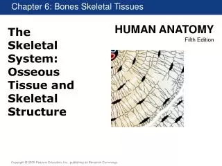
Tissue Structure. Edited by: Jessica Hawley Compiled by Mark Anderson. Classify different tissues by their shape and number Know the four different types of tissues Compare and contrast different functions of tissues. Objectives. How many layers of cells? Single – Simple

E N D
Tissue Structure Edited by: Jessica Hawley Compiled by Mark Anderson
Classify different tissues by their shape and number • Know the four different types of tissues • Compare and contrast different functions of tissues Objectives
How many layers of cells? • Single – Simple • Multiple – Stratified • What is the shape of the cells? • Thin and Flat – Squamous • Square – Cuboidal • Columns – Columnar Classification
Epithelium • Connective Tissue • Nervous Tissue • Muscle Tissue Tissues
Thin layer of tissue that covers all free surfaces of the body • Includes skin and gastrointestinal tract • Simple Epithelium • Located in highly protected areas where maximum absorption is needed • Digestive System • Stratified Epithelium • Located in ares where there is friction with the environment • Skin Epithelium
Provides the structural framework of an animal • All Connective Tissue Includes • Wandering cells • Fixed cells • Ground substance Connective Tissue
Ameboid cells that can freely move within CT • Macrophages • White blood cells • Consume foreign material • Mast cells • Release histamines incase of inflamation • Abundant at the injury site Wandering Cells
Cells that are somewhat anchored within the CT • Fibroblast – determines density of the CT • Primary function is to produce fibrous proteins to reinforce the amorphous structure Fixed Cells
A viscous solution consisting primarliy of proteins linked to carbohydrates • Important in lubricating joints in the form of synovial fluid • Density determined by number of fibrous proteins Ground Substance
Loose – flexible • Dense • Specialized – blood and lymph • Supportive – bones and cartilage Types of connective tissue
Highly porous and flexible • Provides structure for blood vessels and nerves • Highly vascularized • Not very strong Loose CT
Maximum strength with little flexibility • Tendons that are connected to muscle • Two types • Regular – resists force from one directions • Irregular – resists force from multiple directions Dense ct
Adipose tissue • Storage of triglycerides • Highly vascularized • Blood and Lymph • Blood delivers nutrients to tissues • Lymph filters and returns the plasma to the circulatory system • Very little structure Specialized
Cartilage • Hyaline • trachea • Elastic • ear • Fibrocartilage • Between vertebrae • Bone • Spongy • Inside the bone • Compact • Outside the bone Supportive
Hyaline • Densely packed with collagen fibers and provides rigid but flexible features • Elastic • More elastin fibers with some collagen provides maximum flexibility • Fibrocartilage • Extreme form of hyaline cartilage. Very little ground substance Cartilage
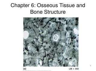
Compact • Very dense and found on the outside of the bones • Spongy • Contains spicules and trabeculae that adds strength Bone
Long bones • Arm and leg bones • Irregular bones • Pelvic bones and vertebrate • Flat bones • Bones of the skull Types of Bones
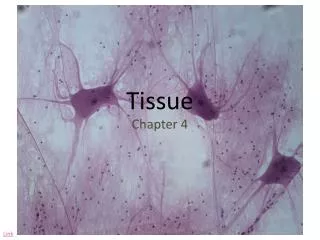
Made of neuron and glial cells and functions to transmit electrical impulses throughout the body • Neuron – nerve cell • Glial cell – support cell that help insulate and support the nerve cell Nervous Tissue
Major features of nerve cells • Axon • Cell Body • Dendrites • Synaptic junctions • Myelin Sheath • Made of Schwann cells Neurons
Functions in locomotion, digestion, breathing, vision, circulation, and other biological processes • Also used as a high protein food source • Makes up 30-40% of total body mass Muscle Tissue
Skeletal – Striated and voluntary • Cardiac – Striated and involuntary • Smooth – Not striated and involuntary Muscle Tissue
Striated and voluntary • Primary muscle type for meat • Multinucleated with nuclei located toward the edge of the cell • Multinucleated cannot reproduce, they can only get larger • Helps with muscle growth Skeletal
Myofiber – muscle fiber/ cell • Myofibril – Contractile apparatus • Sarcolemma – membrane surrounding the myofiber Definitions
A band – darker in color; runs the length of the myosin fiber • I band – lighter in color; only thin filament present • H zone – only thick filament present • Z line – End of the sarcomere where the thin filament is anchored • M line – middle of the sarcomere where thick filament is anchored Sarcomeric structures
Striated and involuntary • Single centrally located nucleus • Varying lengths of the thin filament Cardiac
Not striated, involuntary • Mononucleated, with nucleus in middle of the cell • Tight membrane to membrane junctions for communication between cells Smooth
Classify different tissues by their shape and number • Know the four different types of tissues • Compare and contrast different functions of tissues Objectives