Download
1 / 16
190 likes | 410 Views
Periodontal Ligaments. Introduction. ranges in width from 0.15 to 0.38 mm 0.21 at 11 to 16 years of age 0.18 at 32 to 52 years 0.15 mm at 51 to 67 years. Functions. Supporting the teeth in their sockets Acting as a sensory receptor. Development. Structure. Cells. Extracellular matrix.

E N D
Introduction • ranges in width from 0.15 to 0.38 mm • 0.21 at 11 to 16 years of age • 0.18 at 32 to 52 years • 0.15 mm at 51 to 67 years
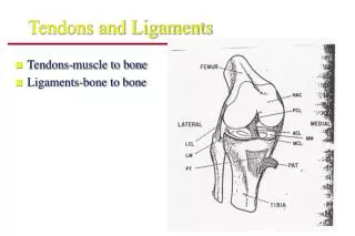
Functions • Supporting the teeth in their sockets • Acting as a sensory receptor
Structure Cells Extracellular matrix Fibroblast Proteins Ground substance Glycosamino- glycan Epithelial cells Collagen fiber bundles Non collagenous protein Glycoproteins Undifferentiated Mesenchymal cells Glycolipids Cementoblasts and Osteoblast
Undifferentiated mesenchymal cells • An important cellular constituent of the PDL
Principal fiber bundles • Alveolar crest group • Horizontal group • Oblique group • Apical group • Interradicular group
Elastic Fibers • There are 3 types of elastic fibers: • Elastin • Oxytalan • Elaunin
Blood Supply • Well vascularized • Perforating arteries • More abundant in posterior than anterior, and in mandible than maxilla • More in gingival and apical third • Venous and lymphatic drain apically
Nerve supply • More in apical area, except for upper incisors, where there is dense distribution in gingival and apical areas.
Nerve Terminations • Free nerve endings (Nociceptor, mechanoreceptor) • Ruffini’s endings (mechanoreceptors) • Coil form • Encapsulated spindle type endings (mechanoreceptors) • Sympathetic Nerve supply