Download
1 / 30
320 likes | 514 Views
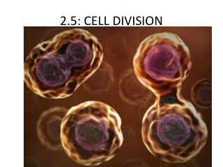
Topic 16 - Eukaryotic Cell Division (Mitosis) & Cell Cycle Control. Similar concept to bacterial division - but more complex. Overview: All DNA duplicated --> increase in cell constituents (organelles, etc.) --> separate the DNA into two copies --> split cell down the

E N D
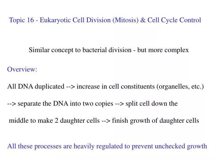
Topic 16 - Eukaryotic Cell Division (Mitosis) & Cell Cycle Control Similar concept to bacterial division - but more complex • Overview: • All DNA duplicated --> increase in cell constituents (organelles, etc.) --> separate the DNA into two copies --> split cell down the middle to make 2 daughter cells --> finish growth of daughter cells • All these processes are heavily regulated to prevent unchecked growth
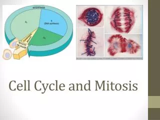
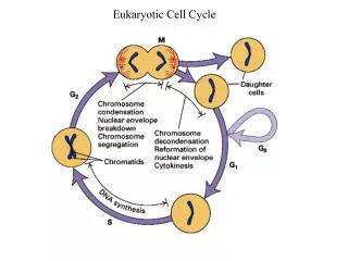
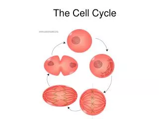
Mitotic Cell Cycle - mitosis & cytokinesis are a small part of cycle fig 12-5
Eukaryotic DNA - amount for whole organism = Genome The large amount of DNA is packaged as Chromatin & then as Chromosomes Chromatin - DNA & protein strands Chromosomes - condensed chromatin - paired as sister chromatids chromatids are joined at centromere fig 12-4 joins the chromatids
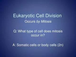
see Fig. 12.4 Each parent donates one of the homologous chromosomes Paired sister chromatids only exist in cells undergoing division
Having two copies of the genome (maternal & paternal) confers an advantage to eukaryotes …. … in this case, surviving malaria …. (sickle cell anemia) one copy of hemoglobin gene is abnormal - but this is actually protective
NOT ON EXAM An aside (but a cool one): Regions of the world with lots of malaria are also regions where sickle cell anemia is common - because it increase malaria survival
Chromatin (duplicated) Aster Centrosomes (with centriole pairs) Early mitotic spindle Centromere Chromosome, consisting of two sister chromatids First stages of mitotic cell division in an animal cell G2 of Interphase Prophase Fig 12-6
Each centrosome contains a pair of centrioles (in animals) The centrosome is a: Microtubule Organizing Centre (MTOC) Plants also have MTOCs, but they lack centrioles See Fig. 6.22 Centrioles 9 of these per centriole
First stages of mitotic cell division in an animal cell Prophase Prometaphase Fig 12-6
Configuration of a chromosome during metaphase Specialized protein plaque See Fig. 12.7
Later stages of mitosis in an animal cell Metaphase Anaphase * Fig 12-6 * not a physical plate - just an area in space like earths equator
Final stages of mitosis & Cytokinesis - in an animal cell Anaphase Telophase & Cytokinesis Fig 12-6
Proposed motor mechanism to move along shortening microtubule Tubulin subunits are removed as the motor passes by so it appears like the chromosome is being pulled to the pole of the cell Chromosomemovement Kinetochore Microtubule Tubulinsubunits Motor protein Chromosome Fig 12-9
(a) Cleavage of an animal cell (SEM) the pinching off of two daughter cells occurs differently if there is a cell wall … Animal cells draw the plasma membrane together in the cell centre using contractile ring of actin & myosin Figure 12.10a 100 m Cleavage furrow Daughter cells Contractile ring ofmicrofilaments
In plants the plasma membrane is partially attached to the cell wall & would tear if pulled away Plants use transport vesicles to deposit membrane & cell wall in a plate between the new daughter cells (b) Cell plate formation in a plant cell (TEM) Figure 12.10b Vesiclesformingcell plate Wall of parent cell 1 m New cell wall Cell plate Daughter cells
Evolution of Cell Division & Mitosis evidence from living eukaryotes that are intermediates between bacteria & higher eukaryotes In Dinoflagellates (protists): the nuclear envelope stays intact. Chromosomes attach to membrane (like bacteria) Microtubules pass thru channels & provide orientation The whole nucleus then divides in two – one for each daughter cell Fig 12-13
Evolution of Cell Division & Mitosis, con't Diatoms (another protist) the Spindle forms within the intact nucleus chromosomes migrate & then the nucleus divides Most animals and plants: Nuclear envelope fragments Fig 12-13
Checkpoints are points in the cell cycle where growth is controlled in eukaryotes controls whether cell divides checks whether cell is ready to divide Fig 12-15
A Checkpoint during mitosis - making sure chromosomes are properly attached Fig 12-8
Most of our cells are non-dividing - they reach the G1 check & stop division, and are said to be in a G0 stage of non-growth Growth Factors - external signals to start dividing can stimulate cells in G0 to start to divide can maintain cell division - used in cell cultures eg. PDGF - stimulates fibroblast division at wound sites eg. EPO - banned at Olympics - stimulates red blood cell production
Cell Culture using growth factors Fig. 12.18
Density and Anchorage dependent cell growth Cancerous Cells “transformed” Fig 12-19
Transformed cells - mutation of genes - defects are passed on..... may make own growth factors or have a defect in a checkpoint Most ‘transformed cells’ are killed by immune system Escapees can grow into Tumours - start from 1 cell. Benign tumour - localized, slow growth - easily treated Malignant tumour - invasive - may change form & chromosomes Cancer! Metastatic - breakaway cells get into circulation & other tissues
Possible spread of untreated Breast Cancer Benign or early malignant malignant metastatic Fig 12-20
Classical approaches to treating cancers – Surgically – but this only works on pre-metastatic cancers Radiation – destroys the ability of all cells to divide Chemotherapy – kills all dividing cells, including non-cancerous ones the holy grail of cancer research is finding a reliable feature of cancer cells that we can target & which our healthy cells don’t have ….
At least in adults such a feature for solid tissue tumors is the need for new blood vessels to provide nutrients and oxygen Research is focusing on how to block the action of growth factors that promote angiogenesis – i.e. new blood vessel growth
Vascular Endothelial Growth Factor (VEGF) inhibitors don't always work NOT ON EXAM blood vessels - red cancer cells - blue normal cells - orange tumour recovers tumour doesn't respond Nature Reviews Cancer8, 592-603 (August 2008)
O2 starved cells send distress signals that cause healthy cells to emit other stimulators of angiogenesis NOT ON EXAM diminished vasculature & oxygen leads to hypoxia (low O2) Nature Reviews Cancer8, 592-603 (August 2008)
Tumour may just adapt by slowing its growth for limited O2 supply NOT ON EXAM blood vessels - red cancer cells - blue normal cells - orange Nature Reviews Cancer8, 592-603 (August 2008)
or metastatic outgrowths may seek new O2 supplies NOT ON EXAM blood vessels - red cancer cells - blue normal cells - orange Nature Reviews Cancer8, 592-603 (August 2008)