Download
1 / 77
780 likes | 982 Views
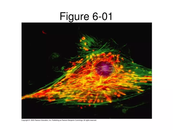
Figure 6-01. Investigating Cell Structure and Function. 1. Cell Theory 2. Microscopy- a. History b. Types 3. Studying cell organelles Cell homogenization b. Cell Fractionation. 10 m. Human height. 1 m. Length of some nerve and muscle cells. Unaided eye. 0.1 m. Chicken egg. 1 cm.

E N D
Investigating Cell Structure and Function 1. Cell Theory 2. Microscopy- a. History b. Types 3. Studying cell organelles • Cell homogenization b. Cell Fractionation
10 m Human height 1 m Length of some nerve and muscle cells Unaided eye 0.1 m Chicken egg 1 cm LE 6-2 Frog egg 1 mm Measurements 1 centimeter (cm) = 10–2 meter (m) = 0.4 inch 1 millimeter (mm) = 10–3 m 1 micrometer (µm) = 10–3 mm = 10–6 m 1 nanometer (nm) = 10–3 µm = 10–9 m 100 µm Most plant and animal cells Light microscope 10 µm Nucleus Most bacteria Mitochondrion 1 µm Electron microscope Smallest bacteria 100 nm Viruses Ribosomes 10 nm Proteins Lipids 1 nm Small molecules Atoms 0.1 nm
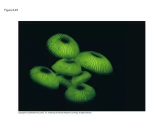
Brightfield (unstained specimen) LE 6-3a 50 µm Brightfield (stained specimen) Phase-contrast
Differential- interference- contrast (Nomarski) Fluorescence LE 6-3b 50 µm Confocal 50 µm
1 µm Scanning electron microscopy (SEM) Cilia LE 6-4 Longitudinal section of cilium Transmission electron microscopy (TEM) Cross section of cilium 1 µm
1 µm Cilia LE 6-4a Scanning electron microscopy (SEM)
Longitudinal section of cilium Cross section of cilium 1 µm LE 6-4b Transmission electron microscopy (TEM)
ENDOPLASMIC RETICULUM (ER Nuclear envelope Flagellum Rough ER Smooth ER NUCLEUS Nucleolus Chromatin Centrosome Plasma membrane LE 6-9a CYTOSKELETON Microfilaments Intermediate filaments Microtubules Ribosomes: Microvilli Golgi apparatus Peroxisome Mitochondrion Lysosome In animal cells but not plant cells: Lysosomes Centrioles Flagella (in some plant sperm)
Inner Life of A Cell • http://www.studiodaily.com/main/searchlist/6850.html
LE 6-5a Homogenization Tissue cells Homogenate Differential centrifugation
1000 g (1000 times the force of gravity) 10 min Supernatant poured into next tube 20,000 g 20 min LE 6-5b 80,000 g 60 min Pellet rich in nuclei and cellular debris 150,000 g 3 hr Pellet rich in mitochondria (and chloro- plasts if cells are from a plant) Pellet rich in “microsomes” (pieces of plasma membranes and cells’ internal membranes) Pellet rich in ribosomes
Cell Structure 1. Basic requirements to be a cell • Cytoplasm • DNA • Ribosome • Cell membrane • Prokaryotic and eukaryotic cells 3. Limitations to cell size • Lower limits • Upper limits-SA/volume ratio
Pili Nucleoid Ribosomes LE 6-6 Plasma membrane Cellwall Bacterial chromosome Capsule 0.5 µm Flagella A typical rod-shaped bacterium A thin section through the bacterium Bacillus coagulans (TEM)
Surface area increases while Total volume remains constant 5 LE 6-7 1 1 Total surface area (height x width x number of sides x number of boxes) 750 6 150 Total volume (height x width x length X number of boxes) 125 125 1 Surface-to-volume ratio (surface area volume) 6 6 1.2
An overview of animal cell structure • Nucleus • Ribosomes • Endomembrane System • RER & SER • Vesicles • Golgi apparatus • Vacuoles • Lysosomes 4. Mitochondria 5. Cytoskeleton
ENDOPLASMIC RETICULUM (ER Nuclear envelope Flagellum Rough ER Smooth ER NUCLEUS Nucleolus Chromatin Centrosome Plasma membrane LE 6-9a CYTOSKELETON Microfilaments Intermediate filaments Microtubules Ribosomes: Microvilli Golgi apparatus Peroxisome Mitochondrion Lysosome In animal cells but not plant cells: Lysosomes Centrioles Flagella (in some plant sperm)
Outside of cell Carbohydrate side chain LE 6-8 Hydrophilic region Inside of cell 0.1 µm Hydrophobic region Hydrophilic region Phospholipid Proteins Structure of the plasma membrane TEM of a plasma membrane
What is contained in the nucleus of a cell? • DNA • Chromosomes • Genes • R-rna • All of the above
Nucleus Nucleus 1 µm Nucleolus Chromatin Nuclear envelope: Inner membrane LE 6-10 Outer membrane Nuclear pore Pore complex Rough ER Surface of nuclear envelope Ribosome 1 µm 0.25 µm Close-up of nuclear envelope Pore complexes (TEM) Nuclear lamina (TEM)
What is the function of ribosomes? • Protein synthesis • DNA synthesis • Intracellular digestion • Transport of proteins outside of the cell
Ribosomes ER Cytosol Endoplasmic reticulum (ER) LE 6-11 Free ribosomes Bound ribosomes Large subunit Small subunit 0.5 µm TEM showing ER and ribosomes Diagram of a ribosome
There is a difference in the make-up of cytoplasmic eukaryotic ribosomes and prokaryotic ribosomes • True • False
Proteins that are secreted from a cell are produced by: • Membrane-bound ribosomes • Free ribosomes
Secreted proteins are carried away from the ER by: • The golgi apparatus • Lysosomes • Mitochondria • vesicles
Smooth ER Nuclear envelope Rough ER LE 6-12 ER lumen Cisternae Ribosomes Transitional ER Transport vesicle 200 nm Rough ER Smooth ER
If a secreted protein needs to be chemically modified after it leaves the ER in a vesicle, it will go to: • A lysosome • Mitochondria • A storage vacuole • The Golgi apparatus
Nucleus Rough ER LE 6-16-3 Smooth ER Nuclear envelope cis Golgi Transport vesicle Plasma membrane trans Golgi
Vesicles from either the ER or the Golgi that contain proteins involved in intracellular digestion fuse to form this cell organelle • Storage vacuole • Mitochondria • Lysosome
Lysosomes are involved in destroying “worn out” cell organelles: • True • False
Up to 5 optional points • You have 3 minutes to write a short answer to this question: • Why is it important that the pH of a lysosome is acidic compared to the cytoplasm of the cell?
1 µm Nucleus LE 6-14a Lysosome Lysosome contains active hydrolytic enzymes Hydrolytic enzymes digest food particles Food vacuole fuses with lysosome Digestive enzymes Plasma membrane Lysosome Digestion Food vacuole Phagocytosis: lysosome digesting food
Lysosome containing two damaged organelles 1 µm Mitochondrion fragment Peroxisome fragment LE 6-14b Hydrolytic enzymes digest organelle components Lysosome fuses with vesicle containing damaged organelle Lysosome Digestion Vesicle containing damaged mitochondrion Autophagy: lysosome breaking down damaged organelle
Malfunctions within a lysosome can cause diseases. • True • False
Vacuoles • Can be formed by endocytosis • May store substances the cell will need later • Can be formed by vesicles joining together • Can be filled with water • All of the above
50 µm Filling vacuole LE 7-14 50 µm Contracting vacuole
This cell organelle has a structure adapted for making ATP during cellular respiration. • Lysosome • Nucleus • Vacuole • Golgi apparatus • mitochondria
Mitochondrion Intermembrane space Outer membrane LE 6-17 Free ribosomes in the mitochondrial matrix Inner membrane Cristae Matrix Mitochondrial DNA 100 nm
The Cytoskeleton • Made up of 3 elements a. Microtubules b. Microfilaments c. Intermediate filaments 2. Functions-diverse including maintaining cells shape; motility; contraction; and organelle movement
Microtubule LE 6-20 Microfilaments 0.25 µm
Centrosome Microtubule LE 6-22 Centrioles 0.25 µm Microtubules Longitudinal section of one centriole Cross section of the other centriole
Cilia and Flagella • Cell Movement
Direction of swimming LE 6-23a Motion of flagella 5 µm
Direction of organism’s movement LE 6-23b Direction of active stroke Direction of recovery stroke Motion of cilia 15 µm
This cell organelle contains 2 compartments separated by a membrane, which is necessary for chemiosmosis to occur • Golgi apparatus • Mitochondria • RER • Lysosome • vacuole
Which of the following statements is/are true? • The cytoskeleton is composed of protein • The cytoskeleton is involved in the segregation of chromosomes during mitosis • The cytoskeleton can reorganize by polymerizing/depolymerizing • A and B • B and C • All of the above
LE 6-24a Microtubules Plasma membrane Basal body 0.5 µm