Download

1 / 16
160 likes | 510 Views
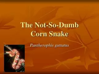
Adenovirus and Cryptosporidium co-infection in a Corn snake ( Elaphe guttata guttata ). Debabrata Mahapatra Department of Infectious Diseases and Pathology. Case History (N08-13). A 13 month old male corn snake was part of the breeding stock of ~2500 mixed Colubrid species.

E N D
Adenovirus and Cryptosporidium co-infection in a Corn snake (Elaphe guttata guttata) Debabrata Mahapatra Department of Infectious Diseases and Pathology Presented at SEVPAC 2008 – Permission granted for use on SEVPAC website only
Case History (N08-13) • A 13 month old male corn snake was part of the breeding stock of ~2500 mixed Colubrid species. • Off-feed about 2 weeks and sharply declining. • Recently moved into a different facility. • Gross findings: No gross lesions. Presented at SEVPAC 2008 – Permission granted for use on SEVPAC website only
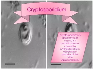
Section of stomach showing mucosal hypertrophy and hyperplasia, mononuclear infiltration in lamina propria and numerous cryptosporidia. (400x) Presented at SEVPAC 2008 – Permission granted for use on SEVPAC website only
EM of sporulating oocysts located in parasitophorous vacuoles (PV) with sporozoites (Sp) and residual body (R) PV PV Sp R Sp Presented at SEVPAC 2008 – Permission granted for use on SEVPAC website only
EM of two schizonts (S) with mature sickle-shaped merozoites (M). An oocyst (O) on the left with feeder organelle (FO) that is separated from the host by a dense band (DB) M S S DB FO M O Presented at SEVPAC 2008 – Permission granted for use on SEVPAC website only
Section of intestine showing villous blunting, expansion of lamina propria with mononuclear cell infiltration. Presented at SEVPAC 2008 – Permission granted for use on SEVPAC website only
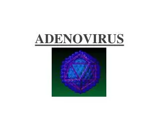
Section of intestine showing prominent large basophilic enterocyte intranuclear inclusion bodies. Presented at SEVPAC 2008 – Permission granted for use on SEVPAC website only
EM of a intranuclear inclusion body (I) with numerous electron dense viral particles (V) within a nucleus (N). I V N Presented at SEVPAC 2008 – Permission granted for use on SEVPAC website only
Morphological diagnosis • Proliferative gastritis, lymphoplasmacytic, diffuse, moderate, with intralesional protozoal parasites consistent with Cryptosporidium sp., stomach. • Enteritis, lymphoplasmacytic, multifocal, mild to moderate, with enterocyte intranuclear inclusions consistent with adenovirus, small intestine. Presented at SEVPAC 2008 – Permission granted for use on SEVPAC website only
Cryptosporidiosis • Cryptosporidium has been reported in over 57 different species of reptiles including: • 40 species of snakes • 15 species of lizards • 2 species of tortoises Of all reptiles, snakes are most severely affected. Lesions are mostly restricted to stomach and intestine with proliferative gastritis and enteritis. Presented at SEVPAC 2008 – Permission granted for use on SEVPAC website only
Cryptosporidiosis • Lizards have a wider range of tissue tropism. Identified in: • In kidneys of Parson’s chameleon. • In kidneys, salivary glands, aural and pharyngeal polyps in green iguana. • Most common species affecting snakes is Cryptosporidium serpentis. • Recently, C. saurophilum has been described in lizards. Presented at SEVPAC 2008 – Permission granted for use on SEVPAC website only
Adenovirus • Adenoviruses are reported in: • 12 different species of reptiles of the orders Crocodilia and Squamata including • Serpentes (snakes) and Sauria (lizards) suborders. • Most of the cases are reported from zoological collection and commercial breeders. Presented at SEVPAC 2008 – Permission granted for use on SEVPAC website only
Adenovirus • Incriminated as the cause of gastroenteritis, hepatitis, nephritis, pneumonia, and encephalitis. • Gastrointestinal lesions have been described in snakes in the families Boidae, Colubridae and Viperidae. • Cytopathologic changes include: • Large eosinophilic to basophilic intranuclear inclusions • Nuclear swelling • Ballooning degeneration • Necrosis of organs Presented at SEVPAC 2008 – Permission granted for use on SEVPAC website only
Conclusion Primary infection with adenovirus might have immunocompromised the snake and predisposed it to secondary infection with cryptosporidia. Presented at SEVPAC 2008 – Permission granted for use on SEVPAC website only
Acknowledgement • Dr. David Taylor • Dr. Lisa Farina • Dr. Mary Reinhard • Dr. Elliot Jacobson’s lab • Lou Ann Miller • UF CVM Histopathology lab members • Residents Presented at SEVPAC 2008 – Permission granted for use on SEVPAC website only
??? Presented at SEVPAC 2008 – Permission granted for use on SEVPAC website only