Download
1 / 29
310 likes | 656 Views
The Limbic System (hippocampus & amygdala). Maggie Donnelly, Anna Edelman, Amanda Lajoie, James Sharpe. Structure & Function Overview. In Latin, limbic means “border” or “edge” since it is the lining of each cerebral hemisphere near the temporal lobe.

E N D
The Limbic System (hippocampus & amygdala) Maggie Donnelly, Anna Edelman, Amanda Lajoie, James Sharpe
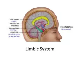
Structure & Function Overview • In Latin, limbic means “border” or “edge” since it is the lining of each cerebral hemisphere near the temporal lobe. • Though there are some structures that are directly associated with the limbic system, many parts that make up the limbic system are part of the cerebral cortex. • Since the limbic system is smaller structure within our cerebral cortex, it does not have the normal 6 layers that make up the neocortex and therefore is commonly referred to as the allocortex or archicortex. • Common disorders that arise from lesions that form on and within structures of the limbic system are autism, various amnesias, Alzheimer’s disease, and Klüver-Bucy syndrome.
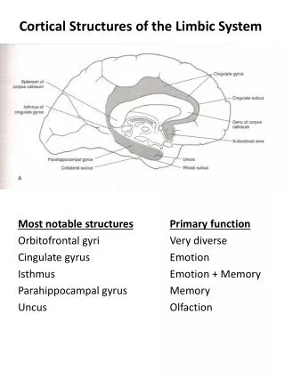
Structures of the Limbic System Amygdala • two masses of neurons on each side of the thalamus, inferior to the hippocampus • signals cortex with stimuli that relate to reward and fear as well as some social functions • also stimulates hippocampus to remember details of a situation Hippocampus • Essential in the formation of long term memories and acts as a converter of short term memories into long term memories Fornix • C shaped bundle of axons • Carries signals from hippocampus to mammillary bodies and septum Mammillary bodies • consist of medial (inner) and lateral (outer) mammillary nuclei • help receive and relay sensory information • Considered part of the hypothalamus
Septal Nuceli (Septum) • composed of medium sized neurons • receive connections from olfactory bulb, hippocampus, amygdala, hypothalamus, midbrain, cingulate gyrus, and thalamus • no relation to smell, pleasure zone in animals Limbic Lobe • Three gyruses: • Parahippocampal, which helps in the formation of spatial memory • Cingulate – automatic functions like heart rate, blood pressure, cognitive processing • Dentate – may contribute to new memories Various publications include other structures.
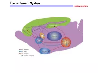
Limbic System Functioning • involved in motivation, emotion, learning, and memory • has influence on endocrine system and the autonomic nervous system • stimulates nucleus accumbens which is part of the ‘pleasure center’ which releases the neurotransmitter dopamine. This is where you get perceive ‘reward-driven’ learning • the basal ganglia, located near the thalamus and hypothalamus. helps in limbic functioning by directing intentional movements receiving input and sending output to motor centers in the brain stem. • The limbic system also has a strong connection to the prefrontal cortex. • One of the ways scientists try to cure severe emotional disorders is my severing the connection, known as a prefrontal lobotomy
main functions in hippocampus Spatial memory • sub-regions in the dentate gyrus in dorsal hippocampus, left hippocampus and parahippocampal region • dorsal hippocampus is important in generating new neurons called adult-born granules which help increase firing in cell networks and contribute to strong memory formations. • The left hippocampus and parahippocampal region helps with recall of spatial memories Learning • high increase in neurons and neural circuits when brain is put through training • these neurons have ‘enhanced excitability’ in the dentate gyrus, which contributes to understanding and retention
main functions of the amygdala Episodic-autobiographical memory (EAM) networks • encoding, storage, and retrieval of spatial memories • neural cues are sent through certain neural networks so that a specific event is re-activated and brought into the working memory Attention and Emotion processes • attention is the ability to focus on some stimuli while ignoring others • Research using EEGs have found that the amygdala helps define a stimulus in order to respond to it accordingly • A researcher named Kheirbek conducted research on how the amygdala contributes to emotional processing and found that neurogenesis (creation) of granule cells was happening in the ventral hippocampus • Though granule cells largely contribute to the strengthening of spatial memory and learning within the hippocampus, they are also seen as a contributor the functioning of the amygdala • A decrease in these cells could result in low emotional functioning and a higher risk for mental illness.
Autism • Disorder of neural development characterized by impaired social interaction and communication, and by restricted and repetitive behavior. • Autism affects information processing in the brain by altering how nerve cells and their synapses connect and organize yet how this occurs is not well understood. • It is one of three recognized disorders in the autism spectrum, with the other two being asperger syndrome and pervasive developmental disorder.
Autism • The diagnostic criteria require that symptoms become apparent before a child is three years old and symptoms are usually noticed in the two first years of a child's life. • Early diagnosis and common signs • Poor eye contact • no smiling or social responsiveness • no response to name • http://www.youtube.com/watch?v=mc1H0aVqn20 • Later indicators include impaired ability to make friends with peers and/or ability to initiate or sustain a conversation with others. Also autism creates restricted patterns of interest that are abnormal in intensity or focus. • Scientists are not certain what causes autism, possibly due to genetics and environmental factors however studies have proven that brain irregularities have an influence
Autism and the Limbic System People with autism often possess an underdeveloped limbic system
Autism and the Limbic System • Underdeveloped amygdala and hippocampus • Problems integrating sensory information • Potential for overactive sense or under-active sense • Neuropsychologist Jocelyne Bachevalier's animal model of autism in monkeys has led to significant evidence toward the limbic system hypothesis. • Social deficits in the monkeys were consistent with the developmental course seen in humans with autism.
Autism Prognosis • There is no known cure and some children recover occasionally either with or without intensive treatment and sometimes not. • Some treatment options include non-medical interventions (behavioral, educational and sensory approaches,communication interventions) and biomedical treatments (modifications in diet, addition of vitamins and minerals, gut treatments and immune system regulations) • Most autistic children can acquire language by age 5 or younger, though a few have developed communication skills in later years. • Although core difficulties tend to persist, symptoms often become less severe with age.
What is Alzheimer's? • Most common form of dementia • Develops differently for every individual • Three stages
What is Alzheimer’s? In Alzheimer's disease, there is an overall shrinkage of brain tissue. The grooves or furrows in the brain, called sulci (plural of sulcus), are noticeably widened and there is shrinkage of the gyri (plural of gyrus), the well-developed folds of the brain's outer layer. In addition, the ventricles, or chambers within the brain that contain cerebrospinal fluid, are noticeably enlarged. In the early stages of Alzheimer's disease, short-term memory begins to fade when the cells in the hippocampus, which is part of the limbic system, degenerate.
Three Stages • The first stage: People with AD the increasing impairment of learning and memory eventually leads to a definitive diagnosis. In a small portion of them, difficulties with language, executive functions, or execution of movements are more prominent than memory problems. • Second Stage: Progressive deterioration eventually hinders independence, with subjects being unable to perform most common activities of daily living. Speech difficulties become evident due to an inability to recall vocabulary, which leads to frequent incorrect word substitutions.
Final Stage During the final stage of AD, the person is completely dependent upon caregivers.Language is reduced to simple phrases or even single words, eventually leading to complete loss of speech.Despite the loss of verbal language abilities, people can often understand and return emotional signals.
What is the hippocampus? • Required for the formation of long- term memories and implicated in maintenance of cognitive maps for navigation. The hippocampus consists of two “horns” that curve back from the amygdalae. It appears to be very important in converting things that are “on one's mind” at the moment (in short-term memory) into things that one will remember for the long run (long-term memory). • The hippocampus is one of the first regions of the brain to suffer damage; memory loss and disorientation are included among the early symptoms.
Alzheimer's and Limbic System • Alzheimer’s disease is a chronic progressive dementia of unknown origin. Early on, patients develop memory impairment which eventually progresses to global intellectual dysfunction. Brain atrophy is more diffuse in Alzheimer’s disease. Senile plaques and neurofibrillary tangles are dispersed throughout the cerebral cortex and basal ganglia, but the hippocampus and amygdala are often severely involved. • Secondary atrophy of the fornix can be seen in chronic cases. Hippocampal atrophy may be a prominent feature on imaging studies, but the finding is nonspecific and the diagnosis of Alzheimer’s disease requires clinical correlation. Volumetric measurements of the amygdala and hippocampus as a unit may be helpful for distinguishing patients with early Alzheimer’s disease.
Shrinking In Hippocampus Area Of Brain Precedes Alzheimer's Disease • The study involved 64 people with Alzheimer's disease, 44 people with mild cognitive impairment, which is the stage of memory problems that precedes Alzheimer's disease, and 34 people with no memory or thinking problems. • MRI scans were performed on all of the participants at the beginning of the study and again an average of a year and a half later. During that time, 23 of the people with mild cognitive impairment had developed Alzheimer's disease, along with three of the healthy participants. • "In people who already have Alzheimer's disease, the loss of nerve cells is more widespread throughout the brain • http://www.youtube.com/watch?v=DKOdEMhO9oM (2:22-2:47)
Amnesia Retrograde Amnesia • loss of memories that formed prior to trauma • temporally graded retrograde amnesia • impairment of memories formed relatively close to when the trauma occurred Anterograde Amnesia • new memories can not be formed after a trauma, but past memories are still intact Both can occur in the same patient Patient H.M.
Amnesia Henry Molaison • Had surgery to cure epilepsy (removed his hippocampus, parahippocampal gyrus, and amygdala) • After surgery had severe anterograde amnesia and temporally graded retrograde amnesia • Able to complete tasks that required short term memory and procedural memory, but not long term episodic memory • suggests that recall from these memory systems may be mediated, at least in part, by different areas of the brain
Anterograde Amnesia and Temporally Graded Retrograde Amnesia for Nonspatial Memory Task after Lesions of Hippocampus and Subiculum Research Article Suggested Explanations of Temporally Graded Retrograde Amnesia (TGRA) • The hippocampus is necessary for memory storage and retrieval for only a limited time after learning • Older memories have a more redundant and spatially distributed representation within the hippocampus than recent memories. TGRA then occurs because a partial lesion of hippocampus is more likely to spare remote memories than a memory acquired recently These ideas have been explored in rats using a learning paradigm based on social transmission of food preference
Anterograde Amnesia and Temporally Graded Retrograde Amnesia for Nonspatial Memory Task after Lesions of Hippocampus and Subiculum Research Article Experiment 1 Purpose: to determine how long the social transmission of food preference can be retained, and whether it changes based on strength of training Experiment 2 Purpose: to determine whether large lesions of the hippocampus and subiculum produce anterograde amnesia for the social food preference task Experiment 3 Purpose: to determine the amnesiatic outcome in rats with large lesions on hippocampus and subiculum made 1, 10, and 30 days after learning
Anterograde Amnesia and Temporally Graded Retrograde Amnesia for Nonspatial Memory Task after Lesions of Hippocampus and Subiculum Research Article Results: • Control group exhibited an 87.4% preference for familiar food and rats with lesions exhibited only a 61.1% preference, which is not greater than chance (exp 2, p< 0.01) • showing that large lesions of the hippocampus and subiculum impair anterograde memory
Anterograde Amnesia and Temporally Graded Retrograde Amnesia for Nonspatial Memory Task after Lesions of Hippocampus and Subiculum Research Article Results continued: • When lesions were made 1 day after training, the operated rats performed at chance (46.2%) and poorer than control animals (74.2%) ( exp 3, p< 0.001) • When lesions were made 10 days after, the operated rats performed numerically worse 54.4 % vs 65.5%,but difference was not significant (exp 3, p> 0.1) • The 30 day lesion group performed the best compared to 10 day and 1 day groups. Also, the 30 day lesion group performed similar to control group 89.3 % vs 79.6% respectively. (exp 3) • the finding that longer training-surgery time improved the performance of the lesion groups provides evidence of temporally graded retrograde amnesia the data suggests then that, with sufficient time, the acquired food preference becomes independent of the hippocampus
multiple choice questions 1. Spatial memory takes place in all of these regions except: a)left hippocampus b)parahippocampal region c)hypothalamus d)dorsal hippocampus 2. Patient H.M suffered from what type of amnesia? a) Post traumatic amnesia b) Temporally graded retrograde amnesia c) anterograde amnesia d) both a and c e) both b and c
multiple choice questions 3. What part of the Limbic System is most affected by Alzhemiers? A. Amlydia B. Hippocampus C. Neither D. All of the Above 4. Children being diagnosed with autism will display the following signs except? a) No response to name b) Poor eye contact c) No smiling or social responsiveness d) Insomnia