Download
1 / 32
320 likes | 465 Views
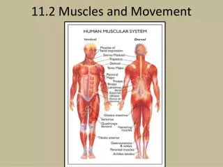
11.2: Muscles & Movement. Which structure is responsible for passing messages directly to effector organs?. 11.2.1 State the roles of ...in human movement. Bones Lever for movement Frame to support body Protect tissue/organs Form blood cells (marrow) Store minerals Ligaments

E N D
Which structure is responsible for passing messages directly to effector organs?
11.2.1 State the roles of ...in human movement. • Bones • Lever for movement • Frame to support body • Protect tissue/organs • Form blood cells (marrow) • Store minerals • Ligaments • Strengthen joint • stabilize bone to bone connections • Muscles • Provide force for movemt by contracting • Shortening length of cells • Occur in antagonistic pairs • Tendons • Attach muscle to bone • Nerves • Sensory ends @ ligaments and muscles • Help prevent over-extension @ joints • Coordinate muscle contraction
11.2.2 Label a diagram of human elbow joint: cartilage, synovial fluid, joint capsule, named bones (humerus, radius, ulna), antagonistic muscles (biceps, triceps)
11.2.2 Label a diagram of human elbow joint: cartilage, synovial fluid, joint capsule, named bones (humerus, radius, ulna), antagonistic muscles (biceps, triceps) http://www.asmi.org/sportsmed/anatomy/elbow.html www.Click4biology.com
11.2.3 Outline the functions of the structures in the human elbow joint. • Hinge joint • Synovial fluid w/in synovial cavity, w/in joint capsule—dense connective tissue, continuous w/bone membranes www.heinemann.co.uk/hotlinks 4242P; weblink 11.1
11.2.5 Describe the structure of striated muscle fibers: myofibrils w/light and dark bands, mitochondria, sarcoplasmic reticulum, nuclei, sarcolemma • Skeletal movement • Muscle fibers = cells • Multinucleate • Plasma membrane = sarcolemma • T tubules = “tunnels” penetrating cell’s interior • Cytoplasm = sarcoplasm • LOTS of stored glycogen (glycosomes) • LOTS of myoglobin (protein) • SR = sarcoplasmic reticulum = smooth ER • Myofibrils = rod-shaped bodies, run length of cell • Lots, packed parallel to each other, lots mitochondria in b/w • Contractile units of the muscle cell; give it striated pattern • “muscle” = 1000s of cells, nerves, vessels
11.2.6 Draw and label a diagram to show structure of a sarcomere: z lines, actin filaments, myosin filaments with heads, and resultant light and dark bands. • Myofibrils: • Sarcomere = unit of movemt • Z lines @ ends • A bands (dArk), length of myosin filaments • H band (narrow) @ middle of A band (myosin only, no actin) w/M line in middle—supporting protein for myosin filaments • I bands (LIght), only actin—no myosin • http://entochem.tamu.edu/musclestruccontractswf/index.html
11.2.6 Draw and label a diagram to show structure of a sarcomere: z lines, actin filaments, myosin filaments with heads, and resultant light and dark bands.
11.2.7 Explain how skeletal muscle contracts, including the release of calcium ions from the SR, the formation of cross-bridges, sliding action of actin and myosin filaments, and use of ATP to break cross bridges and re-set myosin heads. • Sliding filament theory • Actinmyofilaments slide over myosin myofilaments—they don’t shorten! • The sarcomere shortens when they slide over each other • http://3dotstudio.com/zz.html • www.thelifewire.com
Sliding Filament Theory: • Motor neuron carries action potential to NMJ (neuromuscular junction) • Neurotransmitter, acetylcholine, released into gap b/w axon terminal and sarcolemma of muscle fiber • ACh binds to receptors on sarcolemma • Sarcolemma ion channels open, Na+ move through • Generates muscle action potential • Moves along membrane, through T tubules • ACh broken down by acetylcholinesterase to make sure one nerve action potential causes only one muscle action potential
Sliding Filament Theory: 8. Muscle action potential going through T tubules causes release of Ca ions from SR. Flood into sarcoplasm. 9. Ca ions bind to troponin on actin—exposes myosin-binding sites 10. Myosin heads include ATPase which splits ATP and releases energy
Sliding Filament Theory: 11. Myosin heads bind to myosin-binding sites on actin with help of tropomyosin (protein) 12. Myosin-actin crossbridges rotate toward center of sarcomere, producing the “power stroke” 13. ATP binds again to myosin head, resulting in detachment of myosin from actin 14. If no more action potentials, Ca ion level in sarcoplasm falls. Troponin-tropomyosin complex moves to its original position, blocking myosin binding sites...muscle relaxes.
WHOAAAA.... • When you die...Ca++ leak out of SR, bind to troponin...allows actin to slide. But, no ATP produced when you’re dead, so myosin heads can’t detach from actin: RIGOR MORTIS! Lasts ~24 hrs until muscles deteriorate. • Muscle filaments don’t change in length during contraction. • The sarcomere shortens (Z line to Z line)...can see in electromicrographs.
11.2.8 Analyze electron micrographs to find the state of contraction of muscle fibers. • Which is relaxed, which is contracted? • You should also be able to label this!