Download

1 / 33
340 likes | 690 Views
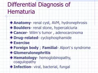
Collecting Duct Carcinoma of Kidney Differential Diagnosis of Neoplasms Involving the Renal Medulla. Merce Jorda, MD, PhD, † and Murugesan Manoharan, MD* ( Pathology Case Reviews 2006;11: 191 – 196). Case Presentation. A 61-year-old woman hematuria and abdominal discomfort.

E N D
Collecting Duct Carcinoma of KidneyDifferential Diagnosis of Neoplasms Involving the Renal Medulla Merce Jorda, MD, PhD,† and Murugesan Manoharan, MD* (Pathology Case Reviews 2006;11: 191–196)
Case Presentation • A 61-year-old woman • hematuria and abdominal discomfort.
macroscopic examination • Left kidney showed a central mass with necrosis measuring 12 cm in greatest dimension • The tumor replaced the entire pyelocaliceal system, invaded the renal vein and the Gerota fascia, and metastasized to the adjacent adrenal gland
Microscopic • this neoplasm was characterized by a predominantly solid and canalicular pattern of growth with papillary areas • Cell size ranged from small cuboidal to large, with eccentric nuclei, distinct nucleoli, and frequent mitoses. • Hobnail cells were also identified
Immunohistochemical stains • high-molecular-weight keratin (HMWK) (+) • E-cadherin (+) • epithelial membrane antigen (EMA) (+) • Carcinoembryonic antigen (CEA) (+) • Ulex europaeus agglutinin-1 UEA-1 (+) • P63 / renal cell carcinoma antigen (RCC) / CD-10 (-)
DISCUSSION • CDC, previously called Bellini duct carcinoma, was first described by Mancilla-Jimenez et al in 1976. • CDC is a rare renal neoplasm that originates from the distal segment of the collecting duct in the renal medulla pyramids known as the collecting ducts of Bellini
DISCUSSION • CDC is thought to arise from the ureteral bud or mesonephros, hence sharing the same origin as urothelial carcinomas. • CDC accounts for 0.6% to 3% of all kidney carcinomas • Of all renal epithelial neoplasms, CDC is the most aggressive, with early metastasis common at the time of clinical presentation
Clinical Features • 2:1 male to female ratio • Mean age is 55 years,wide range (13 to 83) • Association with chronic dialysis • Abdominal pain, flank mass, and hematuria • Imaging studies are usually suggestive of RPUC (pelvic/medullary location)
Clinical Features • Approximately 35% to 40% of patients have metastasis at presentation • Common metastatic sites include regional lymph nodes, adrenal gland, bone, lung, and liver. • More than 75% of patients die with disease within 2 years of diagnosis
Clinical Features • Conventional treatments for renal carcinoma, including radiation, chemotherapy • Immunotherapy are not effective
Gross Pathology • CDC is located in the central region of the kidney and often involves hilar structures. • It ranges in size from 2.5 to 12 cm and is typically firm and gray-white, with infiltrating borders. • It may present as multifocal, probably representing intrarenal metastasis.
Microscopic Characteristics • This neoplasm is characterized by tubular, tubulopapillary, or solid growth patterns with desmoplasia and inflammatory reaction. • Small cyst formations may be present within folding of papillary growth. • Sarcomatoid differentiation as a sign of dedifferentiation has also been described.
Microscopic Characteristics • Mucin production may be present • Cytologically, CDC is composed of eosinophilic or basophilic high-nuclear grade cells with frequent hobnail pattern. • (Papillary RCCs lack hobnail cells)
Immunohistochemistry • HMWK + • UEA-1 + • E-Cad + • CK5/CK6/CK17 - • CD10/15 - • N-Cad - • P63 -
Cytogenetics • Monosomy of chromosome 1 appears to be a constant finding in CDC. • Other alterations described are monosomy of chromosomes 18 and 21, loss of the Y chromosome, and gains of chromosome 7, 12, 17, and 20.
Molecular Alterations • CDCs lose chromosomal arm 1q more frequently than other renal neoplasms, a feature shared with urothelial carcinomas. • Loss of heterozygosity of chromosome 6p is observed in 50% of the CDCs.
Molecular Alterations • Loss of chromosome 3p, frequently seen in conventional RCCs, is a rare event in CDC. Only 8% of CDC demonstrated alteration of the VHL gene (3p25-26). • RCCs have not included CDC due to the rarity of this neoplasm
Differential Diagnosis • The principal differential diagnosis of CDC includes PC, RPUC, and metastatic carcinoma. • Less frequently, other medullary renal neoplasms such as MC and TC may be part of the differential diagnosis
Papillary RCC • a large neoplasm that may invade the renal medulla • histologically characterized by the presence of fibrovascular cores with tumor cells arranged in papillary configuration.
RPUC • RPUC bears little morphologic resemblance to CDC • occasionally extend to collecting ducts of the renal papillae • immunohistochemical markers that may be useful
Medullary carcinoma • MC is a rare neoplasm first distinguished from CDC by Davis et al in 1995 • the strong association of this neoplasm with sickle cell trait is a helpful hint in the differential diagnosis.
Tubulocystic carcinoma • TC, also known as low-grade CDC • Their immunohistochemical profile is similar to CDC • Low nuclear grade and dilated tubules are key diagnostic features of TC
Metastaric carcinoma • Metastatic carcinomas, particularly those of gastrointestinal tract or lung origin, are part of the differential diagnosis. • Immunohistochemistry for CDX-2 and TTF-1 may Be helpful • Frequently multiple, well-circumscribed, and usually not associated with dysplastic changes of the collecting ducts.
CONCLUSION • CDC is a rare and aggressive renal neoplasm that shares biologic characteristics with carcinomas of urothelial origin. • Because of origin in the renal medulla, the differential diagnosis is with other neoplasms that may involve the central area of the kidney. • A correct diagnosis and distinction from other RCCs are imperative since prognosis and treatment modalities are different.