Download
1 / 22
220 likes | 647 Views
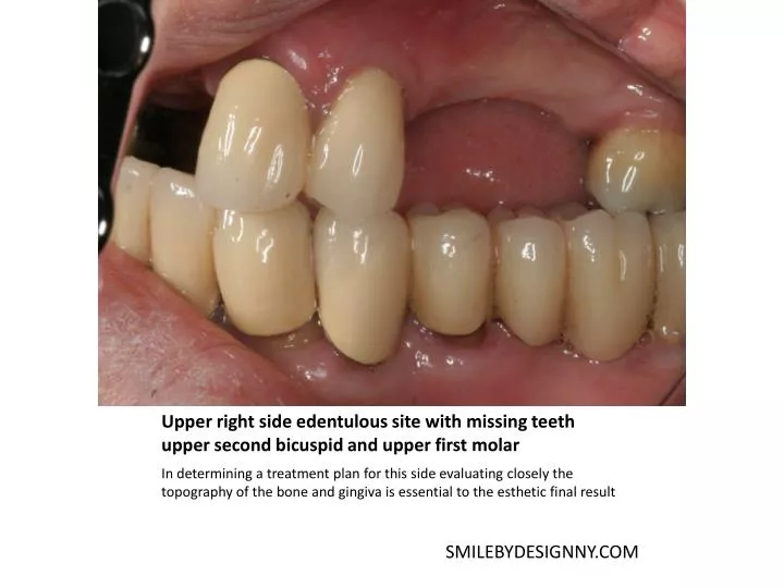
Upper right side edentulous site with missing teeth upper second bicuspid and upper first molar. In determining a treatment plan for this side evaluating closely the topography of the bone and gingiva is essential to the esthetic final result. SMILEBYDESIGNNY.COM. GUIDE PINS FOR SITE #14,15.

E N D
Upper right side edentulous site with missing teeth upper second bicuspid and upper first molar In determining a treatment plan for this side evaluating closely the topography of the bone and gingiva is essential to the esthetic final result SMILEBYDESIGNNY.COM
GUIDE PINS FOR SITE #14,15 Diagnostic casts displays the planning stage for placing 2 implants the upper left side quadrant. SITE #14,15 Angulation of placement ,width of the bone is evaluated SMILEBYDESIGNNY.COM
SURGICAL STENT PLANNING A three dimensional view of the entry point to confirm the parallelism of our procedure. This will be incorporated in the surgical stent once every aspect of the design has been confirmed. SMILEBYDESIGNNY.COM
SITE #14 CT SCAN REPRESENTATION OF UL EDENTULOUS AREA SITE #13 AND 14 FOR TOOTH REPLACEMENT A cross section of the edentulous site shows there is sufficient height of >12mm at site #13 and 10mm height at site #14 to proceed with implant placement. SMILEBYDESIGNNY.COM
SITE #13 CT SCAN OF SITE #13 THE HEIGHT IS APPROX 10MM FROM THE CREST OF THE BONE TO THE BASE OF THE SINUS SMILEBYDESIGNNY.COM
SMILEBYDESIGNNY.COM CLINICAL VIEW OF SITE #13 AND 14 BETWEEN THE UL FIRST BICUSPID AND UL SECOND MOLAR
SURGICAL STENT UR SIDE FOR #13,14 IMPLANT PLACEMENT SMILEBYDESIGNNY.COM
FIRST GUIDE PIN IN OSTEOTOMY FOR INITITIATION PLACEMENT OF IMPLANT SMILEBYDESIGNNY.COM
MEASUREMENT WITH PERIO PROBE OF BONE WIDTH IN SITE #13,14 SMILEBYDESIGNNY.COM
GUIDE PINS #13 14 SMILEBYDESIGNNY.COM
GUIDE PIN PLACED AFTER OSTEOTOMYIN SITE #14 SMILEBYDESIGNNY.COM
8.0MM 10MM 13MM 2.3 MM DRILL IMPL #14 SMILEBYDESIGNNY.COM
3.4/2.8 MM DRILL SMILEBYDESIGNNY.COM
Placed a 4.7x13mm in site #13 and a 4.7x10mm in site #14 SMILEBYDESIGNNY.COM
10.5 MM IMPL BODY 10 MM IMPL 7.5MM 2.5MM 2.5MM #12 #15 SMILEBYDESIGNNY.COM
FINAL SUTURES PLACED SMILEBYDESIGNNY.COM
HEALING AFTER 3 MONTHS IMPLANT PLACEMENT AT SITE #13,14 AND SECOND STAGE UNCOVERING