Download
1 / 24
250 likes | 316 Views
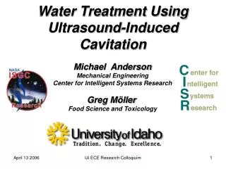
Michiel Postema Professor of E xperimental A coustics KIOGE, Almaty 2012. Using ultrasound to separate oil , gas, and water. INSTITUTT FOR FYSIKK OG TEKNOLOGI. Foam and froth decay. bubble radius ~ mm no -slip interfaces: stable film drainage: very slow. In this talk:.

E N D
Michiel Postema Professor ofExperimentalAcoustics KIOGE, Almaty 2012 Using ultrasoundto separate oil, gas, and water INSTITUTT FOR FYSIKK OG TEKNOLOGI
Foam and froth decay • bubble radius ~ mm • no-slip interfaces: stable • film drainage: very slow
In this talk: • I am going to explain what a foam is; • I am showing how to get rid of foam; • I am going to to show how to force coated bubbles in a liquid to form a foam.
What is a foam? Postema M, et al. Ultrasound-induced microbubble coalescence. UMB 200430(10):1337–1344.
Expanding bubble coalescence Postema M, et al. Ultrasound-inducedencapsulated microbubble phenomena. UMB 2004 30(6):827–840.
Bubble coalescence within 1 microsecond 21 × 21 (µm)² 30 × 30 (µm)² 30 × 30 (µm)² 30 × 30 (µm)² Postema M, et al. Ultrasound-induced microbubble coalescence. UMB 200430(10):1337–1344.
Historic cameras • cameras • 8 – 128 frames • Max. speed (Mfps) • 0.001 (Redlake) • 15 (Brandaris) • 100 (Imacon 468) • 10 – 330 ns exposure • ultrasound • 1 – 10 cycles • 0.5 & 1.7 MHz • P- = 0.04 – 0.85 MPa
Jetting 24 µm bubble Jet 0,33 µs later 60 fl jet volume Postema M et al. IEEE T UFFC 2002(3):c1; Postema M et al. Med Phys 2005 32(12):3707–3711.
Transducer Manufacture Multiple Piezo elementsdiced from the same wafer
Transducer Manufacture Elements lapped down to thickness using slurry of Al2O3in water Ag paint for electrode UV tape as form keeper Very light S-38 microballoon filled epoxy backing
Microbubbles in an ultrasound field 88 × 58 (µm)² area Tx=0.5 MHz, MI=0.09 Equilibrium radius 6 µm +60 kPa Pressure -60 kPa Time 2 µs Postema M et al. Ultrasound-inducedencapsulated microbubble phenomena. UMB 2004 30(6):827–840.
Microbubble resonance frequencies Postema M, Hiltawsky KM, Schmitz G. Ultraschallkontrastmittel – Grundlegende Überlegungen. In: Molecular Imaging – Innovationen und Visionen in der medizinischen Bildgebung; Niederlag W, Lemke HU, Semmler W, Bremer C, Eds. Dresden: Health Academy 2006 (1):131–146.
Fragmentation • 88 × 58 (µm)² area/frame • Tx=0.5 MHz, MI=0.67 • Equilibrium diameter = 4 µm Postema M et al. Presented at Erasmus MC, 2002.
Acoustictabletsmashing Postema M, Smith AJ. Tablet Processing Unit. UK patent application GB0820586.6 2008; international publicationnumber WO/2010/055337.
Sonic cracking • 46 × 30 (µm)² area, solid shell • Tx = 1.7 MHz, PNP 1.5 MPa Postema M, et al. Med Phys2005 32(12):3707-3711.
Radiation forces Kotopoulis S, Postema M. Microfoam formation in a capillary. Ultrasonics 2010 50(2):260–268.
Radiation forces Kotopoulis S, Postema M. Microfoam formation in a capillary. Ultrasonics 2010 50(2):260–268.
Conclusions • We have been able to drive microbubbles through saturated fluids, forcing the bubbles to cluster and form microfoams at equal distances. • These microfoams were then driven out of the fluid. • Ultrasound-assisted separation is a cheap technique that may have applications on a much bigger scale.
Summary of phenomena Postema M, Gilja OH, van Wamel A. CEUS and sonoporation. In: Postema M. Fundamentals of Medical Ultrasonics. London: Spon Press 2011 205–217.
Diffusion Postema M et al. Nitric oxide delivery by ultrasonic cracking: some limitations. Ultrasonics 2006 44:e109–e113.
Elastic bubbles • 40 × 40 (µm)² areas • Tx=0.5 MHz • ≈ 1.1/8 kg s–2 • = r+t+v+s Postema M, de Jong N, Schmitz G. The physics of nanoshelled microbubbles. Biomed Tech 2005 50(S1):748-749.
Phase difference petweenP(t) and R(t) Postema M, Schmitz G. Ultrasonic bubbles in medicine: influence of the shell. UltrasonSonochem 2007 14(4):438–444.