Download
1 / 33
330 likes | 392 Views
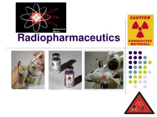
PRINCE SATTAM BIN ABDUL AZIZ UNIVERSITY COLLEGE OF PHARMACY. Nuclear Pharmacy (PHT 433 ). DOSAGE FORMS AND APPLICATION OF RADIOPHARMACEUTICS. Dr. Shahid Jamil. Dosage Forms of Radiopharmaceutics. Solid sources of Radiation :

E N D
PRINCE SATTAM BIN ABDUL AZIZ UNIVERSITY COLLEGE OF PHARMACY Nuclear Pharmacy (PHT 433 ) DOSAGE FORMS AND APPLICATION OF RADIOPHARMACEUTICS • Dr. ShahidJamil L15-16
Dosage Forms of Radiopharmaceutics Solid sources of Radiation: Small Solid sources of Radiation are used for direct implantation into tumours. This has been done for many years with 226Ra and the scope of the technique has been greatly extended by the availability of artificial radionuclides. Removable applicators are used for intracavitary irradiation of the nasopharynx (90Sr), oesophagus 60Co), uterus(137Cs,60Co), bladder (182Ta) and cervix (126Ra). L15-16
Solutions: Including solutions of radiopharmaceuticals for oral administration which is sterile and contain a bacteriostatic or consist of sterile apyrogenic solutions for parenteral use, in single- or multiple- dose vials. Only two radionuclides are widely used in solution form. Thyroid cancer may be treated with 131I, The irradiation of blood-forming tissues by 32P from absorption sites in bone is the method of choice in the treatment of polycythaemia vera, an excessive production of red cells, and may also be useful in the treatment of myeloid and lymphatic leukaemias, which are characterized by excessive white cell production. L15-16
Colloids: Colloidal suspensions of appropriate particle size (10-500 nm) remain located in body cavities or in tissues of low vascularity, this principle is utilized primarily for treatment of the peritoneal and pleural cavities with colloids of gold 198Au and 90Y. These colloids can also be used in the bladder to treatment of diffuse growths. L15-16
Microspheres which are spherical aggregates (0.5-50 μm), generally produced by heat denaturation of biodegradable substances. They are normally suspended in sterile normal parenteral injection and used for, specific capillary blockade. L15-16
Hard gelatin Capsules Either containing some solid in which is absorbed the radiopharmaceutical, or having the radio pharmaceutical evaporated on the inner surface, as in the case of sodium iodide 131 I. Oily liquid radiopharmaceuticals are dispensed in this manner. L15-16
Containers Whether radiopharmaceuticals are supplied as single dose or multidose preparations, the most common container is the 10mL rubber - capped multidose vial. Although glass ampoules are regarded as ideal containers for single dose non-radioactive injections, they are not considered ideal for radiopharmaceuticals due to the potential for radioactive contamination while being opened. L15-16
Labelling of Containers The lable for a radiopharmaceutical should state: • The name of the radiopharmaceutical. • The route of administration. • The activity in the container. • The time and date at which the radiopharmaceutical will have the stated activity. • The volume, if the radiopharmaceutical is in liquid form • The lot number • The expiry date • The name and concentration of any added substances. • Any special instructions such as " Shake well before use". • Any special storage conditions • The name of the supplier • That the preparation is radioactive. L15-16
Medical Applications of Radioisotopes Radioisotopes are used in medicine in two different ways. They may be used as: (1) Radiation sources, or (2) Radioactive tracers. As radiation sources Their principal role is in therapy. The choice of the isotope for a given application is governed by the properties of the radiation required for treatment; type and energy of the radiation and range in tissues. L15-16
As a radioactive tracer The chemical identity and form of the nuclide are most important since the tracer must be isotopic with the element being traced or must otherwise be capable of being incorporated as a part of a particular molecule. The nature of the radiation emitted by a tracer radioisotope is important for its ease of detection. Radioactive tracers are used in medicine principally for diagnostic purposes. L15-16
I- Therapeutic applications of radioisotopes For therapy, isotopes are used as radiation sources, not as tracers. Their therapeutic use is basically justified by the fact that radioactive material, when present in a tissue or organ in sufficient quantity, will produce emanation capable of destroying existing cells and preventing the formation of new tissue. For this reason isotopic therapy is generally applied only to those diseases in which there exists extensive cellular metabolic or to those conditions in which an organ or tissue produces physiological harm through overactivity. L15-16
The radioisotopes as radiation sources for therapy may be used either externally or internally. Where radioisotopes are used as external sources or as sealed sources implanted in a tissue, the dose is terminated by removal of the source. When they are administered internally as an unsealed source, the dose administered to the patient, either in therapy or in diagnosis, cannot be terminated by removal of the source. In therapeutic applications the total dose must be calculated for knowledge of the effective half - life of the isotope, the type and energy of the radiation emitted, and the concentration of the isotope in the tissue. L15-16
A. External Sources 1. Teletherapy units containing 60Co or of 137 Cs have been used for the treatment of lesions. 2. Surface sources for dermatologic and ophthalmologic work consisting of applicators containing pure beta emitters such as 32 P and 90Sr have been used. Where bladder tumors have been treated by infiltration with 32P. 3. Extracorporeal irradiation of blood results in a depletion lymphocytes, thereby producing an alteration in the immunologic response of the individual, 60Co sources were used. L15-16
B- Internal sources Internal sources for radiotherapy for treatment use six different radioisotopes: • Gold Au 198, used as a colloidal gold suspension in the treatment of peritoneal and pleural effusions associated with malignant tumors. 198 Au has also been used in the treatment of prostate and cervical uterine carcinoma and bladder tumors. L15-16
2. Sodium Phosphate P32 may be used in the treatment of polycythemia vera to decrease the rate of formation of the erythrocytes. The isotope is readily distributed to all tissues and is concentrated in those tissues where proliferation is most rapid. Thus cancerous tissues concentrate the greatest amount of the isotope. L15-16
3. Yttrium Y 90 is of special interest because of its 64- hour half - life. It emits a single beta particle no gamma radiation. 90Y has a strong affinity for chelating agents. Chelation With N- hydroxy ethylene diamine triacetic acid (Ed-ol) will cause 90Y to localize in the bone where it can be made to cause predictable hematologic changes. This chelate has been used to treat leukemia and multiple myeloma. L15-16
4. Sodium Iodide I 131has several therapeutic applications. In cases of hyperthyroidism, therapeutic doses of 131I will destroy thyroid tissue by means of radiation produced within the gland. This procedure provides a more desirable mode of therapy than external roentgen-ray treatment since there is less radiation danger to the surrounding tissues. 5. Iodine I125with a half - life of 60 days and an average photon energy of 28 keV is useful for permanent implants for treatment of deep-seated tumors such as those in the chest which are not surgically response. L15-16
Official radiopharmaceuticals used for therapeutic purposes. L15-16
II-Diagnostic Applications of Isotopes For diagnosis, isotopes are used as radioactive tracers and not as radiation sources. When radioisotopes are used for diagnosis, the radiation dose delivered to the patient is maintained at as low a level as possible. This is accomplished by the choice of isotope for the best combination of minimum half-life, minimum retention in the body, and minimum quantity of isotope, which will permit its detection and accurate measurement. L15-16
Radioassary Methods in Medicine Radioisotope studies may be divided into different categories: 1-Activation Analysis 2-Isotope Dilution. 3-Radiometric Analysis. 4-Functional Radioassays. 5- Organ and tumour visualization L15-16
1-Activation Analysis. An analytical technique capable of detecting and measuring certain elements present in a specimen in relatively low concentration if significant amounts of radioactivity are induced by neutron irradiation. L15-16
2-Isotope Dilution. The clinical application of this technique is illustrated by its use for the measurement of blood volume. Radioiodinated human serum albumin injected intravenously; after 10 min injection a blood sample is withdrawn, (a time sufficient to allow adequate mixing of the labeled albumin in the intravascular pool, and not long enough for metabolic activity or leakage into extravascular pools to occur). The blood volume is calculated from the measured decrease in radioactivity of the injected sample upon its dilution by the blood. L15-16
3-Radiometric Analysis. Those radioassay methods which require the use of a standard reagent having a known relationship between chemical concentration and radiologic concentration, i.e., the radioactivity of a specific radioisotope per unit volume, are called radiometric analyses. L15-16
The determination of serum calcium involves the addition of a measured excess of standard 14 C-oxalic acid solution to an aliquot of serum. After precipitation of calcium oxalate the radioactivity of the precipitate is determined. This activity is related to the calcium content of the serum. A variety of similar assays have been developed, including the determination of serum citric acid by use of 82Br. L15-16
4-Functional Radioassays. Radioassays utilizing radioisotopes as aid in measuring the rate of a biological process are called functional radioassays. They can be divided into three categories: i- Rate of isotope transfer. ii-Rate of Isotope Disappearance. iii-Metabolic Processes and/or Isotope Concentration. L15-16
i- Rate of isotope transfer. In these procedures, a labeled substance is injected into one part of the vascular system and the time required for its arrival to another part is determined. This technique has been used widely to determine circulation time, especially in the extremities. 24Na is well suited for this purpose, since: it has a short half-life, is a normal body constituent, is not selectively absorbed by any tissue, and is readily detected. These methods has been used in the treatment of cardiac output. L15-16
ii-Rate of Isotope Disappearance. The rate at which an isotope disappears from a tissue into which it has been injected is a measure of the circulation in that tissue. This test has been used successfully to determine the extent of circulation in tubed skin grafts in plastic surgery. A small amount of an isotope, eg, radiosodium chloride, is injected directly into the tissue. The disappearance rate of the isotope is measured by means of a counter placed directly over the site of injection. L15-16
The RBC destruction mechanism and RBC half-life are measured by means of a disappearance - rate technique. If erythrocytes are labeled in vitro with 51Cr and then reinjected, the rate of the tagged cells can be followed by assay of serial blood samples taken every two or three days for at least two weeks. This study is a valuable aid in the diagnosis of hemolytic anemia. L15-16
iii-Metabolic Processes and/or Isotope Concentration. Most of radioisotope studies are in this category. The concentration of a particular radioisotope in normal or abnormal tissue or in an organ provides data from which the function of the tissue or the metabolic condition of the organ can be evaluated. Several studies of thyroid function can be carried out with the aid of radioactive 131 I as Na 131 I. These studies include: • The rate of deposition of iodine in the gland in vivo. • The total accumulation of iodine in the gland within a specified period of time • The output of thyroid hormone into which radioactive iodine has been incorporated. L15-16
The thyroid gland concentrates inorganic iodide from the blood and converts it to thyroxine (the thyroid hormone), through the action of a peroxidase enzyme. When a thyroid gland is relatively “iodine starved", i.e., receiving no more iodine than is found in the normal diet, the administration of a small dose of radioactive iodine results in a portion of the dose being retained by the thyroid, while the remainder of the radioactive isotope is excreted in the urine. The amount of radioactive iodine retained by the thyroid is an index of thyroid function. L15-16
5. Organ and tumour visualization In recent year scanning have developped rapidly and are now among the most useful tools in diagnostic medicine. By means of scanning, tissues and organs can be visualized and such visualization facilitates the detection of abnormalities in their function. L15-16
In general a scanning technique consists of: a- Administration of a radioactively tagged compound . b- Concentration of the compound in the organ or tissue concerned c- Scanning of the region of the organ to prepare a contour" map of the radioactivity relating the concentration of the radioactivity and its physical location . L15-16
Sites of different radioactivity concentrated in the organs are detected and visible evidence of the pattern of radiation concentration in the area under examination is produced in the form of a photoscan. Dense regions in the scan are indicative of regions of high activity. Organs which have been mapped by such techniques, with particular success, include the lungs, kidneys thyroid, liver, heart, spleen, bone and brain. L15-16