Download
1 / 28
380 likes | 1.1k Views
Hereditary Spherocytosis Ian Roberts. Introduction Causes Symptoms Laboratory Studies Treatment Case Study. Introduction. Red blood cell membrane disorder Spherically shaped red blood cells Most common inherited anaemia in northern European descendants - 1 in 2500

E N D
Introduction • Causes • Symptoms • Laboratory Studies • Treatment • Case Study
Introduction • Red blood cell membrane disorder • Spherically shaped red blood cells • Most common inherited anaemia in northern European descendants - 1 in 2500 • Membrane loss due to defects in membrane proteins: ankyrin, band 3, spectrin and protein 4.2
Causes • Mutations causing defects in membrane proteins • Cause defective or deficient proteins • Affect biconcave shape of red cell
Causes • Mutations causing defects in membrane proteins • Cause defective or deficient proteins • Affect biconcave shape of red cell • Red cells lose shape and become spherical with no flexibility • Unflexible spherocytes get caught up in spleen and phagocytosed
Symptoms • Can range from asymptomatic to severe haemolysis • Anaemia • Jaundice • Fatigue • Splenomegaly
Laboratory Studies • FBC & Retics • Blood film • Bilirubin • LDH • Osmotic Fragility • Dye Binding
Laboratory Studies • Hb • Retics • Bilirubin • LDH • spherocytes on blood film
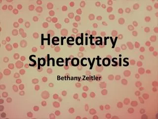
Normal RBC Spherocyte
Laboratory Studies • Hb • Retics • Bilirubin • LDH • spherocytes on blood film • osmotic fragility • dye binding
Osmotic Fragility • Expose RBC to decreasing saline concentrations • As [saline] decreases RBC absorb more • Normal RBC can swell due to flexibility of membrane, therefore are more resistant to low [saline] • Spherocytes are unflexible, therefore cannot absorb more - haemolysis
% Haemolysis ←Decreasing [Saline]
Dye Binding • Flow cytometric method • EDTA anticoagulated peripheral blood incubated with Eosin-5-Maleimide (E5M) • Binds to normal red cell membrane proteins • As dye passes through a laser, it emits light allowing quantitation • HS patients will have reduced dye binding
Normal HStransfused HS
Osmotic Fragility Must be performed within 6 hrs 24hours 3ml blood Unreliable on patients recently transfused Sensitivity 80% (mild cases missed) Abnormal in other causes of spherocytosis (AIHA) Flow Cytometry Reproducible up to 72hrs 1hour 100ul blood Results abnormal after blood transfusion Sensitivity 93% Specificity 99% SEA Ovalocytosis Abnormal in CHAD
Treatment • Phototherapy and/or exchange transfusion in neonates with severe hyperbilirubinaemia • Partial/total splenectomy • Folic acid supplements sustain erythropoiesis
Case Study 1 • Patient A was born in June 2007 • First week Hb dropped slowly, then after 2 weeks sudden drop • Bilirubin was high for first 3 weeks, but decreased after several transfusions and phototherapy • Haematologist suggested tests to confirm HS
Date 20/06/2007 12/06/2007 08/06/2007 05/06/2007 HB 7.3 11.2 14.3 16.7 WBC 16.6 16.6 14.4 14.1 PLT 467 467 296 240 Date 20/06/2007 12/06/2007 08/06/2007 07/06/2007 06/06/2007 05/06/2007 05/06/2007 TBIL 76 110 182 211 238 340 343 • Osmotic fragility test requested on 12/6/07 • Very uncommon test - time consuming - lack of reagents - lack of trained staff
Case Study 1 • Sample sent for Dye binding (very first sample sent) Dye Binding 0.72 units ( 0.80 to 1.20 ) Comments : Reduced dye binding, consistent with diagnosis of hereditary spherocytosis.
Case Study 2 • Patient B was born in November 2009 • First 3 weeks Hb normal, then sudden drop • Bilirubin was high for first 3 weeks, but responded to phototherapy • Haematologist suggested repeating tests 4 months after birth
Date 02/03/2010 14/12/2009 11/12/2009 23/11/2009 22/11/2009 21/11/2009 HB 7.1 8.9 7.7 14.4 16.1 14.5 WBC 7.8 16.5 11.2 9.7 16.6 17.8 PLT 604 582 679 332 245 316 RBC 2.85 2.90 2.45 4.18 4.53 4.14 HCT 0.230 0.260 0.230 0.410 0.450 0.410 Date 02/03/2010 11/12/2009 23/11/2009 22/11/2009 21/11/2009 20/11/2009 TBIL 18 58 164 190 149 103
Case Study • Sample sent for Dye binding • Patient A and Patient B are sisters 13/04/2010 Dye Binding 0.76 units ( 0.80 to 1.20 )
Case Study • Strong family history of HS • Mother has HS – splenectomy in 1991 • Maternal aunt has HS • Maternal Grandmother has HS • ? Paternal history of HS