Download
1 / 49
610 likes | 1.3k Views
Exocytosis and endocytosis. Exocytosis and Endocytosis: Transporting Material Across the Plasma Membrane. Two methods (unique to eukaryotes) for transporting materials across the plasma membrane are Exocytosis, the process by which secretory vesicles release their contents outside the cell

E N D
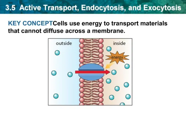
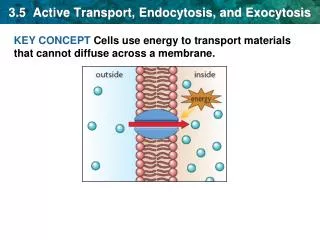
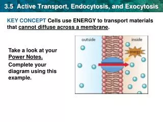
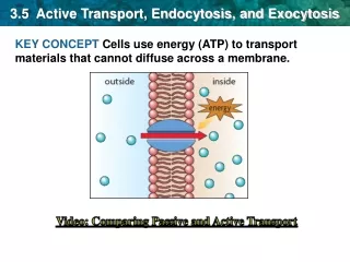
Exocytosis and Endocytosis: Transporting Material Across the Plasma Membrane • Two methods (unique to eukaryotes) for transporting materials across the plasma membrane are • Exocytosis, the process by which secretory vesicles release their contents outside the cell • Endocytosis, the process by which cells internalize external materials
Exocytosis Releases Intracellular Molecules Outside the Cell In exocytosis, proteins in a vesicle are released to the exterior of the cell as the vesicle fuses with the plasma membrane Animal cells secrete hormones, mucus, milk proteins, and digestive enzymes this way Plant and fungal cells secrete enzyme and structural proteins for the cell wall
The process of exocytosis Vesicles containing products for secretion move to the cell surface (1) The membrane of the vesicle fuses with the plasma membrane (2) Fusion with the plasma membrane discharges the contents of the vesicle (3) The membrane of the vesicle becomes part of the cell membrane (4)
Orientation of membrane • When the vesicle fuses with the plasma membrane • The lumenal (inner) membrane of the vesicle becomes part of the outer surface of the plasma membrane • So, glycolipids and glycoproteins that were formed in the ER and Golgi lumens will face the extracellular space
Mechanism of exocytosis The mechanism of the movement of exocytotic vesicles to the cell surface is not clear Evidence points to the involvement of microtubules in vesicle movement Vesicle movement stops when cells are treated with colchicine, a microtubule assembly inhibitor
The Role of Calcium in Triggering Exocytosis • Fusion of regulated secretory vesicles with the plasma membrane is generally triggered by an extracellular signal • In most cases a hormone or neurotransmitter binds receptors on the cell surface and triggers a second messenger inside the cell • A transient elevation in Ca2+ appears to be an essential step in the signaling cascade
Polarized Secretion In many cases, exocytosis of specific proteins is limited to a specific surface of the cell For example, intestinal cells secrete digestive enzymes only on the side of the cell that faces into the intestine This is called polarized secretion
Endocytosis Imports Extracellular Molecules by Forming Vesicles from the Plasma Membrane Most eukaryotic cells carry out one or more forms of endocytosis for uptake of extracellular material A small segment of the plasma membrane folds inward (1) Then it pinches off to form an endocytic vesicle containing ingested substances or particles (2-4)
Membrane flow • Endocytosis and exocytosis have opposite effects, in terms of membrane flow • Exocytosis adds lipids and proteins to the plasma membrane, whereas endocytosis removes them • The steady-state composition of the plasma membrane results from a balance between the two processes
Phagocytosis The ingestion of large particles up to and including whole cells or microorganisms is called phagocytosis For many unicellular organisms it is a means of acquiring food For more complex organisms, it is usually restricted to specialized cells called phagocytes
Phagocytes in immune function In humans, two types of white blood cells use phagocytosis as a means of defense Neutrophils and macrophages engulf and digest foreign materials or invasive microorganisms found in the bloodstream or injured tissues Macrophages are also scavengers, ingesting cellular debris and damaged cells
Phagocytes in the amoeba Phagocytosis is most extensively studied in amoebae, which use it for nutrition Contact with food or other particles triggers the formation of pseudopods, folds of membrane These surround the object and engulf it, forming an intracellular phagocytic vacuole (phagosome)
Phagocytes in the amoeba (continued) The phagosome fuses with a late endosome or matures directly into a lysosome In the lysosome, the engulfed material is digested In the human immune system phagocytes generate toxic amounts of oxidants in the phagosome to kill microorganisms
Receptor-Mediated Endocytosis Cells acquire some substances by receptor-mediated endocytosis (or clathrin-dependent endocytosis) Cells use receptors on the outer cell surface to internalize many macromolecules Mammalian cells can ingest hormones, growth factors, serum proteins, enzymes, cholesterol, antibodies, iron, viruses, bacterial toxins
Low-density lipoproteins Low-density lipoproteins (LDL) are internalized by receptor-mediated endocytosis The internalization of LDL carries cholesterol into cells The study of hypercholesterolemia and connection to heart disease led to the discovery of receptor-mediated endocytosis and a Nobel Prize for Brown and Goldstein
Process of receptor-mediated endocytosis Specific molecules (ligands) bind to their receptors on the outer surface of the cell (1) As the receptor-ligand complexes diffuse laterally they encounter specialized regions called coated pits, sites for collection and internalization of these complexes (2) In a typical mammalian cell, coated pits occupy about 20% of the total surface area
Process of receptor-mediated endocytosis (continued) Accumulation of complexes in the pits triggers the accumulation of additional proteins on the cytosolic surface of the membrane These proteins—adaptor protein, clathrin, dynamin—induce curvature and invagination of the pit (3) Eventually the pit pinches off (4), forming a coated vesicle
Process of receptor-mediated endocytosis (continued) The clathrin coat is released, leaving an uncoated vesicle (5) Coat proteins and dynamin are recycled to the plasma membrane and the uncoated vesicle fuses with an early endosome (6) The process is very rapid and coated pits can be very numerous in cells
Several variations of receptor-mediated endocytosis • Epidermal growth factor undergoes endocytosis and is a signal that stimulates cell division • As EGF receptors are internalized, the cell becomes less responsive to EGF, desensitization • Defective desensitization through failure to endocytose the receptor can lead to excess cell proliferation and possible tumor formation
Variations of receptor-mediated endocytosis • Receptors may be concentrated in coated pits independent of ligand binding • In this case, ligand binding triggers internalization • In another variation (e.g., LDL receptors) receptors are constitutively concentrated and constitutively internalized independent of ligand binding
After internalization Uncoated vesicles fuse with vesicles budding from the TGN to form early endosomes Early endosomes are sites for sorting and recycling of materials brought into the cell Early endosomes continue to acquire lysosomal proteins from the TGN and mature to form late endosomes, which then develop into lysosomes
Recycling plasma membrane receptors Receptors from the plasma membrane are recycled due to acidification of the early endosome The pH gradually lowers as the endosome matures, facilitated by an ATP-dependent proton pump The lower pH dissociates ligand and receptors, allowing receptors to be returned to the membrane
Alternative fates for ligand-receptor complexes • 1. Some complexes are carried to a lysosome for degradation • 2. Some complexes are carried to the TGN, where they enter a variety of pathways • 3. Complexes can also travel by transport vesicles to a different region of the plasma membrane, where they are secreted (transcytosis)
Clathrin-Independent Endocytosis Fluid-phase endocytosis is a type of pinocytosis for nonspecific internalization of extracellular fluid This process does not concentrate the ingested material, and contents are routed to early endosomes It proceeds fairly constantly and compensates for membrane segments added by exocytosis
Coated Vesicles in Cellular Transport Processes Most vesicles in protein and lipid transport are called coated vesicles because of the layers of proteins covering their cytosolic surfaces Coated vesicles are involved in vesicular traffic throughout the endomembrane system, as well as in exocytosis and endocytosis There are several types of coat proteins
Coat proteins The most studied coat proteins are clathrin, COPI, and COPII The type of coat protein on a vesicle helps to determine the destination of the vesicle They also induce curvature needed for the formation of the vesicles and prevent nonspecific fusion of the vesicle with another membrane
Caveolae A recently discovered coat protein is caveolin Caveolin-coated vesicles are caveolae They are a type of lipid raft rich in cholesterol and sphingolipids and may be involved in cholesterol uptake by cells
Clathrin-Coated Vesicles Are Surrounded by Lattices Composed of Clathrin and Adaptor Protein Clathrin-coated vesicles are surrounded by coats made of two multimeric proteins, clathrin and adaptor protein (AP) The shape of clathrin proteins and the way they assemble provides the driving force to induce a flat membrane to form a spherical vesicle The basic unit of clathrin lattices is a triskelion
Structure of a triskelion Each is a multimeric protein composed of three heavy chains and three light chains These radiate from a central vertex, with the light chains associated with the inner half of each “leg” Triskelions assemble into the hexagons and pentagons of the lattice around clathrin-coated pits and vesicles
Adaptor proteins The adaptor protein was identified by its ability to promote assembly of clathrin coats and is sometimes called assembly protein There are four types of AP complexes, each composed of four polypeptides; two of adaptin, one medium chain and one small chain Each polypeptide binds to a different receptor
The Assembly of Clathrin Coats Drives the Formation of Vesicles from the Plasma Membrane and TGN Binding of AP complexes to the plasma membrane and concentration of receptors or receptor-ligand complexes require ATP and GTP The assembly of the clathrin coat appears to provide some of the driving force for vesicle formation
Clathrin coat assembly Initially all clathrin units are hexagonal and form a planar structure As more triskelions are incorporated into the growing lattice, some pentagons are formed The mixture of pentagons and hexagons allows the new coat to curve around the budding vesicle
SNARE Proteins Mediate Fusion Between Vesicles and Target Membranes Once vesicles form, additional proteins ensure delivery to the correct destination The SNARE hypothesis explains this specificity The hypothesis states that sorting and targeting of vesicles involves two families of SNARE (SNAP receptor) proteins
SNARE proteins v-SNAREs (vesicle-SNAP receptors) are found on vesicles t-SNAREs (target-SNAP receptors) are found on target membranes v- and t-SNAREs are complementary molecules that allow recognition between vesicles and their targets