Download
1 / 59
590 likes | 728 Views
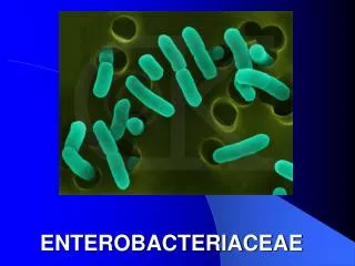
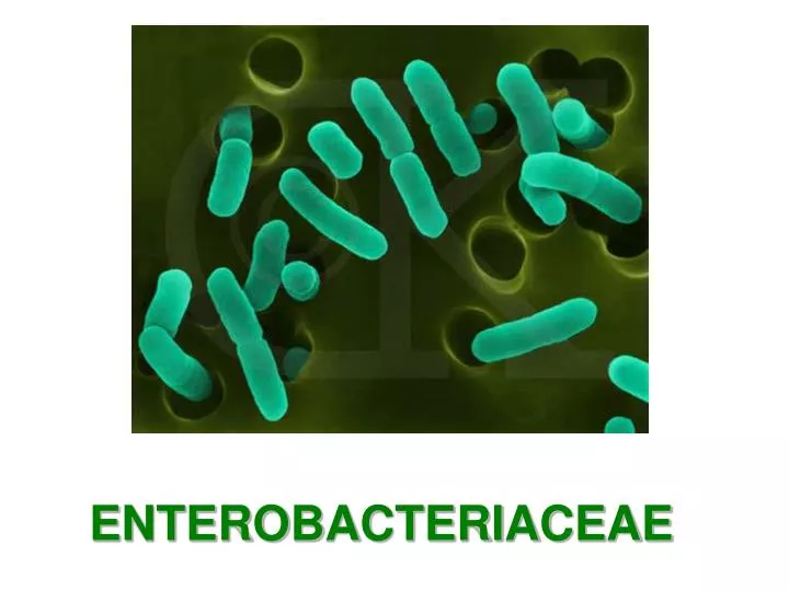
ENTEROBACTERIACEAE. Morphology & Identification. Gram-negative non-spore forming rods. When motile, by peritrichous flagella. Primarily normal flora of gastrointestinal tract. E. coli > Klebsiella > Proteus > Enterobacter Free living, also transient colonizers of skin.

E N D
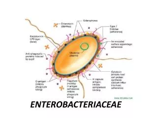
Morphology & Identification • Gram-negative non-spore forming rods. When motile, by peritrichous flagella. • Primarily normal flora of gastrointestinal tract. E. coli>Klebsiella>Proteus>Enterobacter • Free living, also transient colonizers of skin. • Facultative anaerobes: mixed acid fermentation • All ferment glucose; all reduce nitrates to nitrites; all oxidase negative. • Lactose fermentation: normal flora positive and pathogens negative. • Primary isolation media include eosin-methylene-blue (EMB) and MacConkey agar. • Differential selective media for specific organisms including dyes and bile salts. (Salmonella-Shigella (SS) medium, bismuth sulfite media.)
Escherichia Shigella Edwardsiella Salmonella Citrobacter Klebsiella Enterobacter Hafnia Serratia Proteus Providencia Morganella Yersinia Erwinia Pectinobacterium Classification~29 genera, over 100 species.
Antigenic Structure • Most are motile by peritrichous flagella --H antigens. • Capsule –Kantigen (Vifor Salmonella). • Cell envelope (wall) • LPS (endotoxin) –O antigen. • various outer membrane proteins. • Pili - various antigen types, some encoded by plasmids
鞭毛抗原(H) K或Vi抗原 菌体抗原(O)
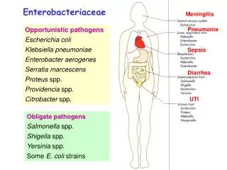
Enterobacteriaceae- Opportunistic diseases Citrobacter Enterobacter Escherichia Hafnia Morganella Providencia Serratia • septicemia, • pneumonia, • meningitis • urinary tract infections
Enterobacteriaceae:gastrointestinal diseases • Escherichia coli • Salmonella • Shigella • Yersinia entercolitica
Enterobacteriaceae • community acquired • otherwise healthy people • Klebsiella pneumoniae • respiratory diseases • prominent capsule • urinary tract infection • fecal contamination • E. coli • Proteus • urease (degrades urea) • alkaline urine
Escherichia coli • Toxins: two types of enterotoxin; Shiga-type toxin; Enteroaggregative ST-like toxin; Hemolysins; Endotoxin • Type III secretion system • Adhesions –colonization factors ; both pili or fimbriae ;non-fimbrial factors involved in attachment. There are at least 21 different types of adhesions. • Virulence factors that protect the bacteria from host defenses: Capsule/Iron capturing ability (enterochelin) • Outer membrane proteins
E. coli fimbriae Type 1 mannose P • galactose • glycolipids • glycoproteins
E.coli-urinary tract infection Is the leading cause of urinary tract infections which can lead to acute cystitis (bladder infection) and pyelonephritis (kidney infection).
E.coli-Meningitis and Sepsis • Neonatal meningitis – is the leading cause of neonatal meningitis and septicemia with a high mortality rate. Usually caused by strains with the K1 capsular antigen.
Enteropathogenic E. coli • fever • infantdiarrhea • vomiting • nausea • non-bloody stools • Destruction of surface microvilli • loose attachment mediated by bundle forming pili (Bfp); • Stimulation of intracellular calcium level; • rearrangement of intracellular actin,
Enterotoxigenic E. coli • A watery diarrhea, nausea, abdominal cramps and low-grade fever for 1-5 days. • Travellers diarrhea and diarrhea in children in developing countries • Transmission is via contaminated food or water.
Enterotoxigenic E. coli • diarrhea like cholera • milder • nursery travellers diarrhea • caused by LT, ST, or LT/ST.
Enterotoxigenic E. coli • Heat labile toxin • like choleragen • Adenyl cyclase activated • cyclic AMP • secretion water/ions • Heat stable toxin • Guanylate cyclase activated • cyclic GMP • uptake water/ions
E.coli-Enteroinvasive (EIEC) • The organism attaches to the intestinal mucosa via pili • Outermembrane proteins are involved in direct penetration, invasion of the intestinal cells, and destruction of the intestinal mucosa. • There is lateral movement of the organism from one cell to adjacent cells. • Symptoms include fever,severe abdominal cramps, malaise, and watery diarrhea followed by scanty stools containing blood, mucous, and pus. • resembles shigellosis
Enteroinvasive E. coli (EIEC) • Dysentery • resembles shigellosis • elder children and adult diarrhea
E.coli-c. Enteropathogenic (EPEC) • Malaiseand low grade feverdiarrhea, vomiting, nausea, non-bloody stools • Bundle forming pili are involved in attachment to the intestinal mucosa. • This leads to changes in signal transduction in the cells, effacement of the microvilli, and to intimate attachment via a non-fimbrial adhesion called intimin. • This is a problem mainly in hospitalized infants and in day care centers.
E.coli-d. Enterohemorrhagic (EHEC) • Hemorrhagic • bloody, copious diarrhea • few leukocytes • afebrile • hemolytic-uremic syndrome • hemolytic anemia • thrombocytopenia (low platelets) • kidney failure
Transmission electron micrograph Enterohemorrhagic E. coli • Usually O157:H7
Enterohemorrhagic E. coli • Vero toxin • “shiga-like” • Hemolysins • younger than 5 years old,causing hemorrhagic colitis
Enteroaggregative E. coli 肠集聚型大肠杆菌 • a cause of persistent, watery diarrhea with vomiting and dehydration in infants. • That is autoagglutination in a ‘stacked brick’ arrangement. • the bacteria adheres to the intestinal mucosa and elaborates enterotoxins (enteroaggregative heat-stable toxin, EAST). • The result is mucosal damage, secretion of large amounts of mucus, and a secretory diarrhea.
E.coli-Enteroaggregative (EAggEC) • Mucous associated autoagglutinins cause aggregation of the bacteria at the cell surface and result in the formation of a mucous biofilm. • The organisms attach via pili and liberate a cytotoxin distinct from, but similar to the ST and LT enterotoxins liberated by ETEC. • Symptoms incluse watery diarrhea, vomiting, dehydration and occasional abdominal pain.
Sanitary significance • Totoal bacterial number: number of bacteria contained per ml or gm of the sample; the standard of drinking water is less than 100. • Coliform bacteria index: the number of coliform bacteria detected out per 1000 ml sample; the standard of drinking water is less than 3
Shigella • S. flexneri, S. boydii, S. sonnei, S. dysenteriae • bacillary dysentery • shigellosis • bloody feces • intestinal pain • pus
Genral features • Pili. • Most strains can not ferment lactose; S. sonnei can slowly_ ferment lactose. • According to O antigen, 4 groups • Easily causing drug-resistence.
Shigellosis • within 2-3 days • epithelial cell damage
Shiga toxin • enterotoxic • cytotoxic • inhibits protein synthesis • lysing 28S rRNA
Shigella attachment and penetration • Within 2-3 days • Epithelial cell damage
Clinical significance • man only "reservoir" • mostly young children • fecal to oral contact • children to adults • transmitted by adult food handlers • unwashed hands
Clinical significance • The infective dose required to cause infection is very low (10-200 organisms). • There is an incubation of 1-7 days followed by fever, cramping, abdominal pain, and watery diarrhea (due to the toxin)for 1-3 days. • This may be followed by frequent, scant stools with blood, mucous, and pus (due to invasion of intestinal mucosa). • Is is rare for the organism to disseminate. • The severity of the disease depends upon the species one is infected with. S. dysenteria is the most pathogenic followed by S. flexneri, S. sonnei and S. boydii.
Immunity • SIgA.
Diagnosis of Shigella infection • Specimen:stool. • Culture and Identification • Quick immunological methods: • Immunofluorescent “ball” test; • Coagglutination.
Prevention • streptomycin dependent (SD) dysentery vaccine.
Treating shigellosis • manage dehydration • patients respond to antibiotics , Problem of drug-resistance • disease duration diminished
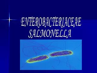
Salmonella • Salmonellosis may present as one of several syndromes including gastroenteritis, enteric (typhoid) fever or septicemia.
The antigenic structures of salmonellae used in serologic typing
Salmonella • 2000 antigenic "types” • genetically single species • S. enterica • disease category • S. enteritidis • many serotypes • S. cholerae-suis • S. typhi
Virulence factors • Endotoxin – may play a role in intracellular survival • Capsule (for S. typhi and some strains of S. paratyphi) • Adhesions – both fimbrial and non-fimbrial • Type III secretion systems and effector molecules – 2 different systems may be found: • One type is involved in promoting entry into intestinal epithelial cells • The other type is involved in the ability of Salmonella to survive inside macrophages • Outer membrane proteins - involved in the ability of Salmonella to survive inside macrophages • Flagella – help bacteria to move through intestinal mucous • Enterotoxin - may be involved in gastroenteritis • Iron capturing ability
Enteric or typhoid fever • Enteric or typhoid fever occurs when the bacteria leave the intestine and multiply within cells of the reticuloendothelial system. • The bacteria then re-enter the intestine, causing gastrointestinal symptoms. • Typhoid fever has a 10-14 day incubation period and may last for several weeks. • Salmonella typhi is the most common species isolated from this salmonellosis. • Human reservoir:carrier state common • Contaminated food:water supply • Poor sanitary conditions
Typhoid • Septicemia • -occurs 10-14 days • lasts 7 days • gall bladder • shedding, weeks • acute phase, gastroenteritis gastrointenteritis
伤寒和付伤寒的致病过程 胆囊---肠道---粪排菌/肠 壁淋巴组织 肾-----尿 肝脾-----肿大 骨髓------受抑制 皮肤----血栓出血--玫瑰疹 伤寒和付伤寒沙门菌 小肠上部粘膜 固有层淋巴结 进入血液 再次进入血液 肠系膜淋巴结 第一次菌血症 第二次菌血症