Download
1 / 23
340 likes | 748 Views
Structure and Specificity of Caspases . Ning Chen. Caspases. Caspase stands for c ysteine asp artate-specific prote ase . Caspases have the characteristics of high specificity for substrates containing Asp, and use a Cys for catalyzing peptide bond cleavage.

E N D
Structure and Specificity of Caspases Ning Chen
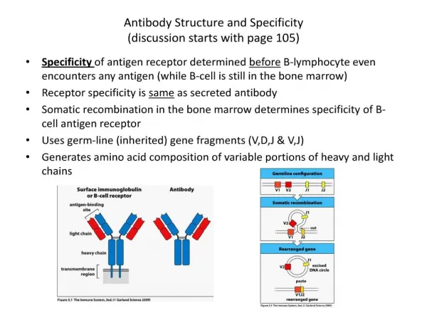
Caspases • Caspase stands for cysteine aspartate-specific protease. • Caspases have the characteristics of high specificity for substrates containing Asp, and use a Cys for catalyzing peptide bond cleavage. • Caspases are the major executioners in apoptosis. • Caspases, a unique family of cysteine protease, execute apoptosis, which was first documented in 1993 under the condition of the discovery that CED3 plays a central role in the programmed cell death in worm Caenorhabditis elegans. Stennicke and Salvesen, Cell Death and Differentiation (1999) 6, 1054 – 1059 Friedlander,N Engl J Med (2003) 348, 14
Apoptosis • Apoptosis is also known as programmed cell death, which is an important regulator of growth, development, defense, and homeostasis in multi-cellular organisms. • Apoptosis study has become a major research in the biomedical science. • More than 15000 papers published annually on this topic, is over 2% of the papers published in the life sciences.
Apoptosis and diseases • Too much apoptosis: neurodegenerative diseases, such as Parkinson’s diseases, Alzheimer’s diseases, spinal muscular atrophy. • Too little apoptosis: Cancers (virus infection or DNA mutations). • Too little apoptosis: autoimmune diseases, such as diabetes type I, encephalomyelitis. • Some cardiovascular diseases and liver diseases are also related to apoptosis. • Many toxins and other cellular stresses can also trigger apoptosis, such as oxidative stress, alcohol.
Apoptosis Pathways Prior and Salvesen, Biochem. J. (2004) 384, 201–232
Group I Group II Group III • Caspase Family Denault and Salvesen, Chem. Rev. 2002, 102, 4489-4499
Caspase Structure Prior and Salvesen, Biochem. J. (2004) 384, 201–232
Caspase Activation Concha and Meguid, Current Medicinal Chemistry (2002)9, 713-726
Active caspase tetramer Donepudi and Grütter, Biophysical Chemistry (2002) 101-102, 145-153
Active Caspase Chang and Yang, Microbiology and Molecular Biology Review, 2000, 821–846
Caspase Binding Site Stennicke and Salvesen, Cell Death and Differentiation (1999) 6, 1054 - 1059
Major Interactions with Caspase Active Sites Nicholson, Cell Death and Differentiation (1999) 6, 1028 -1042
Caspase Substrate Specificity Nicholson, Cell Death and Differentiation (1999) 6, 1028 -1042
Caspase Proteolytic Specificity Nicholson, Cell Death and Differentiation (1999) 6, 1028 -1042
Role for S5 Site in Substrate Recognition Fang et al., J. Mol. Biol. (2006) 360, 654–666
Cleavage site CFP YFP CFP YFP cleavage + Fluorescence Resonance Energy Transfer (FRET) Luo, et al., Biochem and Biophys Res Comm., 304 (2003): 217
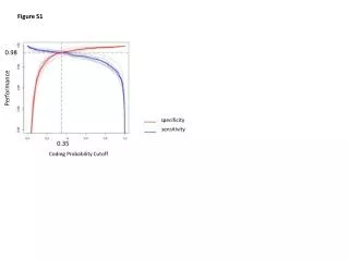
P2 Absorbance and/or Fluorescence Change engineering cleavage sites protease cleavage P3 P1 EGFP EGFP-based protease sensors Design of EGFP-based Caspase Sensors
Opportunities • Based on caspase properties, determining caspase activity can be used for investigating the mechanisms of diseases. • Based on the selectivity and specificity in caspase substrate, rational design caspase triggers and inhibitors for treating diseases related to caspase activity. • Determining caspase activity in real time in cells or in vivo can be used for tracking the process of diseases and effects of treatments or for screening caspase inhibitors in drug development.
Challenges • Some oncoproteins can activate caspases and induce apoptosis, however, transfected cells can still survive, therefore, the apoptosis pathways still need to be further investigated and completed in transfected cells or in vivo. • Although several classes of selectively potent reversible and irreversible caspase inhibitors have been identified, it is still a difficult task to develop selective small-molecular nonpeptide inhibitors for pharmaceutical uses. • Caspase activity determination in cells or in vivo in real time is still a big challenge.
References • Stennicke, H.R. and G.S. Salvesen, Catalytic properties of the caspases. Cell Death Differ, 1999. 6(11): p. 1054-9. • Friedlander, R.M., Apoptosis and caspases in neurodegenerative diseases. N Engl J Med, 2003. 348(14): p. 1365-75. • Fuentes-Prior, P. and G.S. Salvesen, The protein structures that shape caspase activity, specificity, activation and inhibition. Biochem J, 2004. 384(Pt 2): p. 201-32. • Denault, J.B. and G.S. Salvesen, Caspases: keys in the ignition of cell death. Chem Rev, 2002. 102(12): p. 4489-500. • Concha, N.O. and S.S. Abdel-Meguid, Controlling apoptosis by inhibition of caspases. Curr Med Chem, 2002. 9(6): p. 713-26. • Donepudi, M. and M.G. Grutter, Structure and zymogen activation of caspases. Biophys Chem, 2002. 101-102: p. 145-53. • Chang, H.Y. and X. Yang, Proteases for cell suicide: functions and regulation of caspases. Microbiol Mol Biol Rev, 2000. 64(4): p. 821-46. • Nicholson, D.W., Caspase structure, proteolytic substrates, and function during apoptotic cell death. Cell Death Differ, 1999. 6(11): p. 1028-42. • Fang, B., et al., Structural and kinetic analysis of caspase-3 reveals role for s5 binding site in substrate recognition. J Mol Biol, 2006. 360(3): p. 654-66. • Kang, P.M. and S. Izumo, Apoptosis in heart: basic mechanisms and implications in cardiovascular diseases. Trends Mol Med, 2003. 9(4): p. 177-82.
References • Ghavami, S., et al., Apoptosis in liver diseases--detection and therapeutic applications. Med Sci Monit, 2005. 11(11): p. RA337-45. • Shi, Y., Caspase activation, inhibition, and reactivation: a mechanistic view. Protein Sci, 2004. 13(8): p. 1979-87. • Grutter, M.G., Caspases: key players in programmed cell death. Curr Opin Struct Biol, 2000. 10(6): p. 649-55. • Shi, Y., Mechanisms of caspase activation and inhibition during apoptosis. Mol Cell, 2002. 9(3): p. 459-70. • Riedl, S.J. and Y. Shi, Molecular mechanisms of caspase regulation during apoptosis. Nat Rev Mol Cell Biol, 2004. 5(11): p. 897-907.
Thank you! Questions are welcome!