Download
1 / 15
160 likes | 474 Views
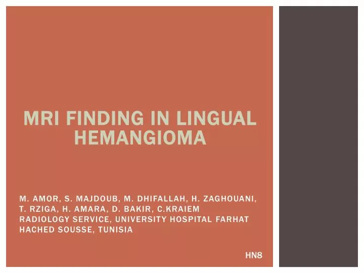
M. AMOR, S. MAJDOUB, M. DHIFALLAH, H. ZAGHOUANI, T. RZIGA, H. AMARA, D. BAKIR , C.KRAIEM Radiology service, University Hospital Farhat Hached Sousse, Tunisia. MRI FINDING IN LINGUAL HEMANGIOMA. HN8. Objectives. To describe the plain and enhanced MRI findings of lingual hemangioma .

E N D
M. AMOR, S. MAJDOUB, M. DHIFALLAH, H. ZAGHOUANI, T. RZIGA, H. AMARA, D. BAKIR, C.KRAIEM Radiology service, University Hospital FarhatHached Sousse, Tunisia MRI FINDING IN LINGUAL HEMANGIOMA HN8
Objectives • To describe the plain and enhanced MRI findings of lingual hemangioma. • To discuss the importance of contrast medium in the differential diagnosis of high intensity lesions of the tongue on T2 weighted images.
Materials and methods • The clinical records and MR images of two patients affected by a lingual hemangioma were reviewed. • Patients presented with a palpable submucosal bluish-red soft mass in the tongue.
Materials and methods • MRI examinations were performed. Plain and enhanced SE (spin echo) T1 weighted and FSE (fast spin echo) T2 weighted images were acquired in axial, sagittal and coronal planes. • Axial T1 weighted images were also obtained before and after intravenous injection of paramagnetic contrast medium.
Results • The two cases MR imaging shows the lesion as a solid mass with iso or slightly high signal intensity to muscle on T1-weighted images (figure 1) and heterogeneous signal intensity on T2-weighted images (figure 2 and 3).
Figure 2: axial fat sat T2- weighted Figure 1: axial fat sat T1-weighted Figure 3: sagittal fat sat T2-weighted
Results • PostcontrastT1-weighted imaging commonly demonstrates prominent enhancement (figure 4, 5 and 6). • Due to the presence of multiple low signal intensity vessels with rapidly flowing blood, the hemangiomas have a typical serpentine appearance.
Figure 5: sagittal fat sat T1-weighted+gado Figure 6: coronal fat sat T1-weighted+gado Figure 4: axial fat sat T1-weighted+gado
Discussion • Hemangiomais the most common tumor of infancy. • The basis of the cellular features described by Mulliken and Glowacki in 1982, vascular lesions may be classified as either hemangioma or vascular malformation. • They proposed that the term ‘hemangioma’ should be limited to those lesions that show increased mitotic activity.
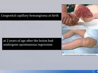
DISCUSSION • Most hemangiomas are present at birth, but some do not manifest clinically until early childhood. They may regress spontaneously due to internal bleeding, thrombosis, or organization. • The most common sites of oral occurrence are the lip, buccal mucosa, tongue, and palate. • Lingual hemangiomas may remain indolent or may produce obstructive symptoms or alarming hemorrhage
Discussion • Most lingual tumors present as mucosal changes and tongue being superficially located and easily accessed, these can be diagnosed without imaging analysis. • However, the characteristic and extent of lesions situated at deep portion of tongue, such as its base or submucosal lesions can be recognized only on cross- sectional CT scan or MRI.
DISCUSSION • MR imaging shows the lesion as a solid mass with iso- or slightly high signal intensity to muscle on T1-weighted images and heterogeneous signal intensity on T2-weighted images. • PostcontrastT1-weighted imaging commonly demonstrates prominent enhancement.
DISCUSSION • Due to the presence of multiple low signal intensity vessels with rapidly flowing blood. • Some hemangiomas have a typical serpentine appearance. The conspicuity of these signal void areas increases with tumor size.
DISCUSSION • Small lesions can be excised with impunity. • Large lesions, if excised, could result in significant functional disability. This is why several modalities of less invasive treatment have recently been advocated (Argon laser, YAG laser, or both to avoid functional disability caused by tissue loss). • There have been reports of treatment with superselective embolization using polyvinyl alcohol foam and absorbable gelatin sponge particulates.
Conclusion • Haemangiomasare among the most common neoplasms encountered in the paediatric age group. • Knowledge of MRI findings of lingual hemangioma is useful for differential diagnosis with other high intensity lingual lesions on T2 weighted images. • This discrimination is achievable using iv paramagnetic contrast medium.