Download
1 / 1
10 likes | 127 Views
Case of the Day – Monday Musculoskeletal. Smith SE 1 , Davis KW 2 University of Maryland School of Medicine, Baltimore, MD University of Wisconsin, Madison, Wisconsin. History: 42 year old male with bilateral hip pain and history of renal transplants.

E N D
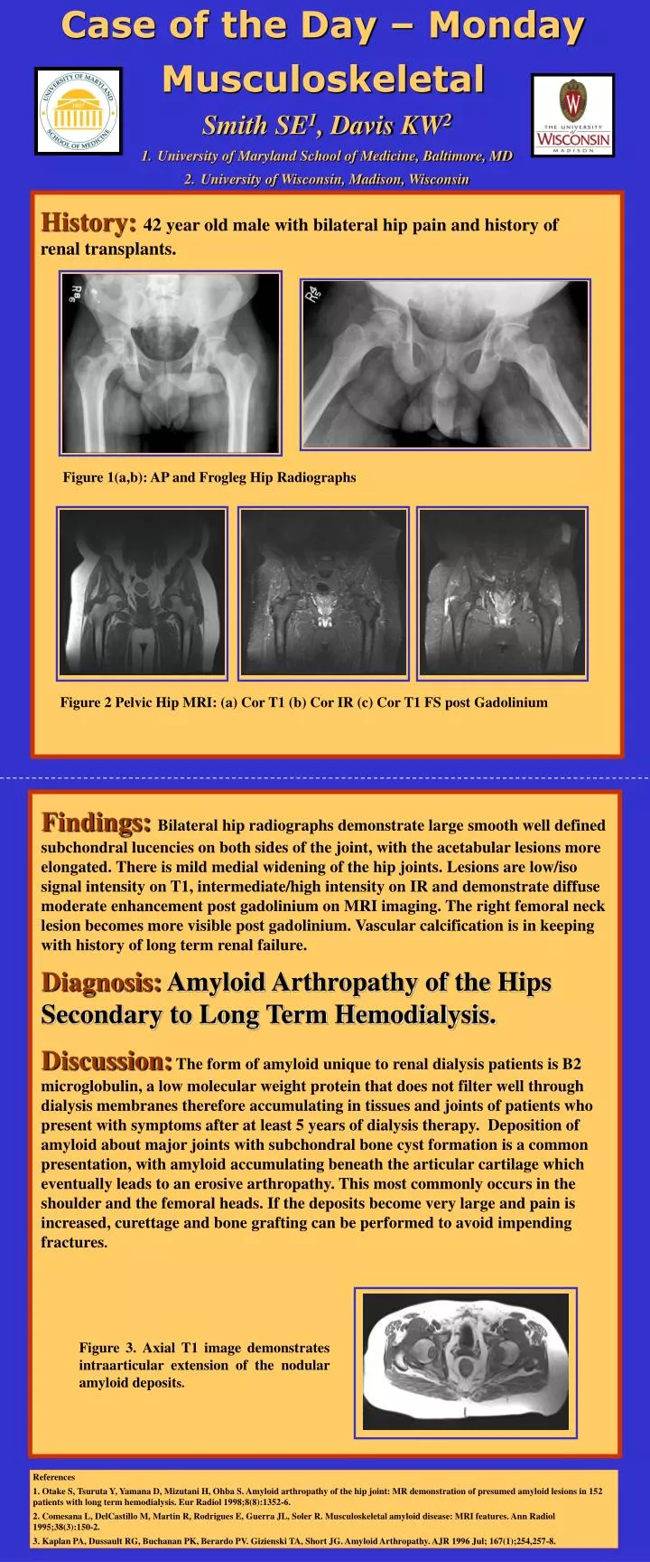
Case of the Day – Monday Musculoskeletal • Smith SE1, Davis KW2 • University of Maryland School of Medicine, Baltimore, MD • University of Wisconsin, Madison, Wisconsin History:42 year old male with bilateral hip pain and history of renal transplants. Figure 1(a,b): AP and Frogleg Hip Radiographs Figure 2 Pelvic Hip MRI: (a) Cor T1 (b) Cor IR (c) Cor T1 FS post Gadolinium Findings: Bilateral hip radiographs demonstrate large smooth well defined subchondral lucencies on both sides of the joint, with the acetabular lesions more elongated. There is mild medial widening of the hip joints. Lesions are low/iso signal intensity on T1, intermediate/high intensity on IR and demonstrate diffuse moderate enhancement post gadolinium on MRI imaging. The right femoral neck lesion becomes more visible post gadolinium.Vascular calcification is in keeping with history of long term renal failure. Diagnosis: Amyloid Arthropathy of the Hips Secondary to Long Term Hemodialysis. Discussion:The form of amyloid unique to renal dialysis patients is B2 microglobulin, a low molecular weight protein that does not filter well through dialysis membranes therefore accumulating in tissues and joints of patients who present with symptoms after at least 5 years of dialysis therapy. Deposition of amyloid about major joints with subchondral bone cyst formation is a common presentation, with amyloid accumulating beneath the articular cartilage which eventually leads to an erosive arthropathy. Thismost commonly occurs in the shoulder and the femoral heads. If the deposits become very large and pain is increased, curettage and bone grafting can be performed to avoid impending fractures. Figure 3. Axial T1 image demonstrates intraarticular extension of the nodular amyloid deposits. References 1. Otake S, Tsuruta Y, Yamana D, Mizutani H, Ohba S. Amyloid arthropathy of the hip joint: MR demonstration of presumed amyloid lesions in 152 patients with long term hemodialysis. Eur Radiol 1998;8(8):1352-6. 2. Comesana L, DelCastillo M, Martin R, Rodrigues E, Guerra JL, Soler R. Musculoskeletal amyloid disease: MRI features. Ann Radiol 1995;38(3):150-2. 3. Kaplan PA, Dussault RG, Buchanan PK, Berardo PV. Gizienski TA, Short JG. Amyloid Arthropathy. AJR 1996 Jul; 167(1);254,257-8.