Download
1 / 68
700 likes | 908 Views
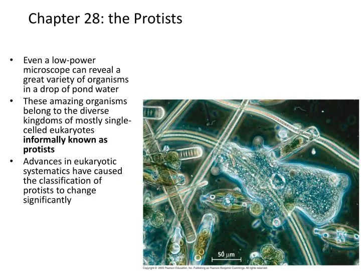
Chapter 28: the Protists. Even a low-power microscope can reveal a great variety of organisms in a drop of pond water These amazing organisms belong to the diverse kingdoms of mostly single-celled eukaryotes informally known as protists

E N D
Chapter 28: the Protists • Even a low-power microscope can reveal a great variety of organisms in a drop of pond water • These amazing organisms belong to the diverse kingdoms of mostly single-celled eukaryotes informally known as protists • Advances in eukaryotic systematics have caused the classification of protists to change significantly
Kingdom Protista?? • now part of the superkingdomEukaryota • eukaryotes = true nucleus • evolution of a nucleus for the genetic information • evolution of membrane-bound organelles • diverse group of single and colonial forms informally known as The Protists • but Kingdom Protista really doesn’t exist anymore – too polyphyletic • probably arose from more than one prokaryotic group • 7 to 45 species recognized depending on zoologist • some as small as prokaryotes • molecular analysis has discovered many commonalities that make them Protists
Protists • include groups that are photoautotrophs, heterotrophs and mixotrophs • mixotrophs = combine photosynthesis and heterotrophic nutrition • divide the protists into three categories: • 1. Photosynthetic – plant-like algae • 2. Ingestive – animal-like protozoans • 3. Absorptive – fungus-like
Cellular Anatomy • most are unicellular • but the cellular composition is extremely complex • unicellular protists carry out similar functions to multi-cellular eukaryotes with their organ systems • do so using subcellular organelles • many of these organelles are seen in higher organisms • other organelles are not found in the typical multicellular eukaryote • contractile vacuoles for osmoregulation
Protists and Eukaryotic Evolution • Many components of the eukaryotic animal and plant cell were derived from protists • diversity of protists has its origins in endosymbiosis • process where a unicellular organism engulfs another cell – become endosymbionts and eventually a new organelle
Protists and Eukaryotic Evolution • early evolution – ingestion of a photosynthetic cyanobacteria through primary endosymbiosis by a primitive eukaryote • eventual development into the plastids of the photosynthetic red and green algae • Red and green algae also underwent secondary endosymbiosis • they themselves were ingested by another primitive eukaryotic cell to become eventual plastids of the protists listed below in the figure Plastid Dinoflagellates Secondary endosymbiosis Apicomplexans Red algae Cyanobacterium Primary endosymbiosis Stramenopiles Heterotrophic eukaryote Secondary endosymbiosis Plastid Euglenids Secondary endosymbiosis Green algae Chlorarachniophytes
The 5 Supergroups of Eukaryotes • 1. Excavata • 2. Chromalveolata • the alveolates and stramenophiles • 3. Rhizaria • 4. Archaeplastida • contains green algae and land plants • 5. Unikonta • slime molds, entamoebas, fungi and animals
Eukaryotic Phylogenetic Tree Excavata Chromalveolata Rhizaria Unikonta Archaeplastida Rhodophyta Euglenozoa Parabasala Diplomonadida Radiolaria Cercozoa Fungi Animalia Plantae Chlorophyta Charophyta Stramenopila Amoebozoa (Opisthokonta) Alveolata (Viridiplantae) Choanoflagellates Dinoflagellates Chlorarachniophytes Euglenids Ciliates Charophyceans Plants Diplomonads Oomycetes Foraminiferans Radiolarians Gymnamoebas Entamoebas Fungi Metazoans Red algae Diatoms Cellular slime molds Chlorophytes Parabasalids Brown algae Kinetoplastids Apicomplexans Golden algae Plasmodial slime molds Ancestral eukaryote
Clade: Excavata • A. Diplomonads • B. Parabasilids • C. Euglenozoans
Clade: Excavata • Diplomonads & Parabasilids • protists in these two clades lack plastids (no photosynthesis) • mitochondria do not have DNA or the enzymes for the citric acid cycle or proteins for the electron transport chain
Clade: Excavata • A. Diplomonads • two equal-sized nuclei and multiple flagella • flagella is very different from prokaryotic flagella • have modified mitochondria = mitosomes • many are parasites giardia intestinalis
LE 28-5b Flagella Undulating membrane 5 µm Trichomonas vaginalis, a parabasalid (colorized SEM) • B. Parabasalids • also have reduced/modified mitochondria = hydrogenosomes • include the protists called trichomonads– Trichomonasvaginalis • mobility through an undulating membrane in addition to flagella
C. Euglenozoans • belong to a diverse clade – includes heterotrophs, photosynthetic autotrophs and parasites • considered a photosynthetic protist similar to algae • like algae – the photosynthetic protists have chlorophyll a and b in chloroplasts • distinguishing feature – a rod with either a spiral or crystalline structure inside each of their flagella • divided into the groups: • 1. the Kinetoplastids • 2. the Euglenoids
1. Kinetoplastids - Trypanosomes • used to be called the zoomastigophores • defined by a single, large mitochondrion that contains an organized mass of DNA = kinetoplast • free-living forms in freshwater, marine and soil – feed on the prokaryotes in these ecosystems • some are parasites of animals, plants and other protists • Trypanosomagambienese– sleeping sickness (neurological disease) & Chagas’ disease (congestive heart failure) in humans
Kinetoplastids: Trypanosoma Life cycle -cycles between the tsetse fly and the human-different forms of the trypanosome depending on what host and where it is in the host fly injects the trypanosome multiplication in the human host – e.g. in the blood bit by fly and transfer multiplication in the fly’s gut and then in the salivary gland
2. Euglenoids – The Euglena • unicellular protist • most are autotrophic • several chloroplasts with chlorophyll a and b and carotenoid pigments • some can also be mixotrophic – photosynthetic in sunlight, engulfs prey in absence of sunlight • main characteristic - two flagella that emerge from a “pocket” structure • at the pocket is a large contractile vacuole that connects to the outside • continuously collects water from the cell and returns it to the outside – regulates osmotic pressure • two flagella arise at this reservoir • only one emerges from the canal and actively beats for locomotion • used to be classified as the Class Phytomastigophorea
2. Euglenoids • inside the plasma membrane is a structure called the pellicle • articulated strips of protein lying side by side • elastic enough to enable turning and flexing of the protist • but rigid enough to prevent major changes in shape • eyespot (stigma) - near the flagella • functions as a pigment shield allowing only certain wavelengths of light to strike the light detector • light detector (photoreceptor) – detects the filtered light and results in movement toward the light direction • probably developed in order to maximize its photosynthetic potential • used to be classified as the Class Phytomastigophorea
Clade: Chromalveolata • originated more than a billion years ago when their ancestor ingested a photosynthetic red algae (via secondary endosymbiosis) • plastids within these protists have red algae origins (DNA analysis) • divided into two major groups: • 1. Alveolates • 2. Stramenophiles
Clade: Chromalveolata • A. Alveolates: • 1. Dinoflagellates • 2. Apicomplexans • 3. Ciliates • B. Stramenophiles • 1. Diatoms • 2. Golden Algae • 3. Brown Algae • 4. Oomycetes
Chromalveolata - A. Alveolates • characterized by membrane-bound sacs called alveoli • just under the plasma membrane • function unknown • 1. Dinoflagellates – move through flagellar action • 2. Apicomplexans- parasites • 3. Ciliates – move through ciliary action
LE 28-10 Flagella 3 µm Alveolates: 1. Dinoflagellates • several thousand species • “dinos” = whirling • components of both marine and freshwater phytoplankton • possess characteristic shapes – reinforced by internal plates of cellulose that become encrusted with silica - act as “armor” • some can be heterotrophic (phagocytic) • most are autotrophic with well-formed plastids for photosynthesis • possess mitochondria with tubular cristae (similar to animals) • two flagellae– located in perpendicular grooves in these plates • one groove is transverse =cingulum– propels the dinoflagellate forward and causes it to spin • other groove is longitudinal = sulcus – acts as the rudder
capable of proliferating explosively – “blooms” • “red tide”(carotenoid pigments found in the plastids) - produce a toxin that kills off invertebrates • some can be bioluminescent – ATP driven reaction that creates a glow at night • may be a defense mechanism
Alveolates: 2. Apicomplexans • nearly all are animal parasites • spread through the formation of tiny infectious cells = sporozoites • named because one end (apex) contains a complex of organelles specialized for penetrating host tissues and cells • have a non-photosynthetic plastid = apicoplast which has many functions including the synthesis of fatty acids for its membranes • life cycle – includes sexual and asexual stages • requires more than one host to complete
Alveolates: 2. Apicomplexans • best known is the Plasmodium – causes malaria • rivals tuberculosis as the leading cause of human death by infectious disease • can be reduced by insecticides that kill the Anopheles mosquito (DDT) and by drugs that kill the Plasmodium (quinine based drugs) • vaccines hard to develop – Plasmodium lives inside the RBC (hidden) • carriers of sickle cell anemia gene – resistant to malaria
LE 28-11 Inside mosquito Inside human Merozoite Sporozoites (n) Liver Liver cell Oocyst Apex MEIOSIS Merozoite (n) Red blood cell 0.5 µm Zygote (2n) Red blood cells FERTILIZATION Key Gametes Haploid (n) Gametocytes (n) Diploid (2n) Plasmodium Life Cycle • 1. infected Anopheles mosquito bites a person injecting its sporozoites (n) • 2. sporozoites enter the liver and undergo division to become merozoites (n) • merozoites enter RBCs by using their apical complex • 3. the merozoites asexually divide to make more • some go on to infect more RBCs • 4. other merozoites develop into gametocytes • 5. gametocytes picked up by a new mosquito • 6. gametes form and fertilization takes place in the mosquito’s digestive tract • the fertilized cell = zygote • 7. an oocystdevelops from the zygote and adheres to the wall of the mosquito’s gut • produces more sporozoites • these are delivered to a new human host when the mosquito bites another human
Alveolates: 3. Ciliates - Paramecium • use of cilia to move and feed • cilia may completely cover the protist or may cluster in a few rows or tufts • distinguished by the presence of two types of nuclei: macronucleus (large) and micronucleus (small) • may have one or more of each type • macronucleus – contains dozens of copies of the genome • control the everyday functions of the ciliate • micronucleus – function in reproduction • exchanged between two ciliates during conjugation
LE 28-12 Paramecium FEEDING, WASTE REMOVAL, AND WATER BALANCE Paramecium feeds mainly on bacteria. Rows of cilia along a funnel-shaped oral groove move food into the cell mouth, where the food is engulfed into food vacuoles by phagocytosis. Paramecium, like other freshwater protists, constantly takes in water by osmosis from the hypotonic environment. Bladderlike contractile vacuoles accumulate excess water from radial canals and periodically expel it through the plasma membrane. Contractile vacuole Oral groove Cell mouth • freshwater protist – constantly takes on water from its hypotonic environment • they contain contractile vacuoles for the regulation of osmotic pressure – accumulate excess water via radial canals and then expel it through the plasma membrane back into the environment Thousands of cilia cover the surface of Paramecium. Food vacuoles combine with lysosomes. As the food is digested, the vacuoles follow a looping path through the cell. 50 µm Micronucleus The undigested contents of food vacuoles are released when the vacuoles fuse with a specialized region of the plasma membrane that functions as an anal pore. Macronucleus
Paramecium FEEDING, WASTE REMOVAL, AND WATER BALANCE • cilia participate in movement • but also gather food and move it toward the oral groove which holds the cell mouth at the bottom • food is then engulfed into a food vacuole via phagocytosis • food vacuoles combine with lysosomes containing digestive enzymes • undigested food particles are carried to the opposite end of the cell as the cell mouth • fuse with the plasma membrane in a specific region – acts as an “anal pore” Paramecium feeds mainly on bacteria. Rows of cilia along a funnel-shaped oral groove move food into the cell mouth, where the food is engulfed into food vacuoles by phagocytosis. Paramecium, like other freshwater protists, constantly takes in water by osmosis from the hypotonic environment. Bladderlike contractile vacuoles accumulate excess water from radial canals and periodically expel it through the plasma membrane. Contractile vacuole Oral groove Cell mouth Thousands of cilia cover the surface of Paramecium. Food vacuoles combine with lysosomes. As the food is digested, the vacuoles follow a looping path through the cell. 50 µm Micronucleus The undigested contents of food vacuoles are released when the vacuoles fuse with a specialized region of the plasma membrane that functions as an anal pore. Macronucleus
Paramecium • asexual reproduction – through binary fission • sexual reproduction involves conjugation • 1. two compatible mating strains align side by side and partially fuse • 2. meiosis of their micronuclei produces a total of 4 haploid micronuclei in each cell • 3. three micronuclei in each disintegrate & the remaining micronuclei in each divides by mitosis- resulting in 2 micronuclei in each paramecium • 4. the cells swap one of their micronuclei – genetic recombination • 5. the cells separate CONJUGATION AND REPRODUCTION Meiosis of micronuclei produces four haploid micronuclei in each cell. Three micronuclei in each cell disintegrate. The remaining micro-nucleus in each cell divides by mitosis. Two cells of compatible mating strains align side by side and partially fuse. Compatible mates The cells swap one micronucleus. Macronucleus MEIOSIS Haploid micronucleus Diploid micronucleus Diploid micronucleus MICRONUCLEAR FUSION The cells separate. Two rounds of cytokinesis partition one maccronucleus and one macronucleus into each of four daughter cells. Micronuclei fuse, forming a diploid micronucleus. The original macronucleus disintegrates. Four micronuclei become macronuclei, while the other four remain micronuclei. Three rounds of mitosis without cytokinesis produce eight micronuclei. Key Conjugation Reproduction
Paramecium • 6. the two micronuclei in each cell fuse to produce a diploid nuclei • 7. three round of mitosis without fission results in 8 micronuclei in each paramecium • 8. the original macronuclei disintegrates and 4 micronuclei become 4 macronuclei to replace it – leaves 4 micronuclei • 9. two rounds of binary fission now happen results in 4 daughter cells • 10. the micronuclei (4) and macronuclei (4) then partition into the four daughter cells – each paramecium ends up with 1 micronuclei and 1 macronuclei CONJUGATION AND REPRODUCTION Meiosis of micronuclei produces four haploid micronuclei in each cell. Three micronuclei in each cell disintegrate. The remaining micro-nucleus in each cell divides by mitosis. Two cells of compatible mating strains align side by side and partially fuse. Compatible mates The cells swap one micronucleus. Macronucleus MEIOSIS Haploid micronucleus Diploid micronucleus Diploid micronucleus MICRONUCLEAR FUSION The cells separate. Two rounds of cytokinesis partition one maccronucleus and one macronucleus into each of four daughter cells. Micronuclei fuse, forming a diploid micronucleus. The original macronucleus disintegrates. Four micronuclei become macronuclei, while the other four remain micronuclei. Three rounds of mitosis without cytokinesis produce eight micronuclei. Key Conjugation Reproduction
Got all that?? CONJUGATION AND REPRODUCTION Meiosis of micronuclei produces four haploid micronuclei in each cell. Three micronuclei in each cell disintegrate. The remaining micro-nucleus in each cell divides by mitosis. Two cells of compatible mating strains align side by side and partially fuse. Compatible mates The cells swap one micronucleus. Macronucleus -partially fuse -1 micronuclei becomes 4 via meiosis (haploid) -3 disappear -1 micronuclei becomes 2 via mitosis -paramecia “swap” 1 micronuclei and separate -fuse 2 micronuclei into 1 (diploid) -2 micronuclei become 8 (mitosis/no cytokinesis) -macronuclei disappears -so 4 of the 8 micronuclei develop into 4 macronuclei -4 of the micronuclei stay micronuclei -2 rounds binary fission 4 daughter paramecia -each daughter cell gets a macronuclei and a micronuclei MEIOSIS Haploid micronucleus Diploid micronucleus Diploid micronucleus MICRONUCLEAR FUSION The cells separate. Two rounds of cytokinesis partition one macronucleus and one macronucleus into each of four daughter cells. Micronuclei fuse, forming a diploid micronucleus. The original macronucleus disintegrates. Four micronuclei become macronuclei, while the other four remain micronuclei. Three rounds of mitosis without cytokinesis produce eight micronuclei. Key Conjugation Reproduction
Chromalveolata - B. Stramenophiles • stramen = “straw”; pilos – “hair” • comprised of several groups of heterotrophs and several groups of phototrophs(considered to be algae) • flagella are said to be “hairy” – have numerous hair-like projections along the length • this hairy flagellum is paired with a smooth flagellum • 1. oomycetes – water molds • 2. bacillariophytes - diatoms • 3. chrysophytes – golden algae • 4. charophyceans – brown algae Hairy flagellum Smooth flagellum 5 µm
What is Algae?? • photsyntheticprotists • algae= eukaryotic organism with chlorophyll a pigments that carry out oxygen-producing photosynthesis • study of algae = phycology • no longer any formal classification schemes • algae are scattered across many phyla = polyphyletic • BUT They differ from plants – lack a well-organized vascular system and they have a simple reproductive system • occur most often in water • fresh and marine – may be suspended as planktonic organisms or attached to the bottom (benthic)
Algae: Photosynthetic Protists • algae frequently confused with plankton • plankton = free-floating microscopic aquatic organisms • phytoplankton – made up of algae and small plants • zooplankton – non-photosynthetic protists and animals • classical algae are now grouped together with the plants - Phyla Chlorophyta • some are a separate lineage - known as red algae • Phylum Rhodophyta • some are grouped with the stramenophiles- yellow and brown algae • Phyla Chrysophyta and Phaeophyta
Algae: Photosynthetic Protists • important properties that classify them: • 1. cell wall composition – rigid cell wall • some have an outer membrane outside the wall – similar to the bacterial capsule • 2. the form in which food is stored • 3. chlorophyll molecules and accessory pigments (carotenoids) • chloroplasts are found in membrane-bound sacs (thylakoids) for the light-reactions of photosynthesis • 4. flagella number and location of their insertion into the cell • flagella are used for locomotion • 5 morphology of the cells and/or body • comprised of a vegetative body = thallus
Algae: Photosynthetic Protists • important properties that classify them: • 6. habitat: marine or freshwater • unicellular, colonial, filamentous, membranous, blade-like or tubular • 7. reproductive structures: reproduction is asexual or sexual • asexual – seen in unicellular forms • sexual – generation of eggs by oogoniaor sperm by antheridia • 8. mitochondria cristae structure: tubular, disc or plate-like (lamellar)
Stramenophiles: 1. Oomycetes: Water molds • oomycete = “egg fungus” • water molds, white rusts and downeymildews • white rusts and downey mildews live as parasites on land plants • e.g. Potato blight - Phytophthorainfestans water mold
Stramenophiles: 1. Oomycetes: Water molds • used to be considered fungi – have multinucleate filaments called hyphae that resemble those seen in fungi • but the oomycetes have cell walls made of cellulose (fungus – chitin) and the diploid condition predominates (reduced in fungi) • molecular data also cannot confirm fungal origins • similarities are an example of convergent evolution • do not carry out photosynthesis – non-autotrophic • acquire nutrients as decomposers – grow as cottony masses on dead animals and algae = heterotrophic water mold
life cycle: can alternate between asexual and sexual forms • a zoospore develops via mitosis into a hyphae • the zoospore is biflagellated with one smooth flagella and the other “hairy” • so it is a stramenophile • these hyphae will develop zoosporangia at their tips - produce zoospores asexually (i.e. mitosis) • but hyphae can also develop sexual structures that produce gametes via meiosis Oogonium Germ tube Egg nucleus (n) Cyst Antheridial hypha with sperm nuclei (n) MEIOSIS ASEXUAL REPRODUCTION Zoospore (2n) FERTILIZATION Zygote germination Zygotes (2n) SEXUAL REPRODUCTION Zoosporangium (2n) Key Haploid (n) Diploid (2n)
life cycle: sexual • one region of the hyphae undergoes meiosis to produce egg nuclei (n) within a structure called an oogonium • other branches can develop sperm nuclei (n) via meiosis – contained within an antheridialhyphae • these antheridialhyphae grow and “hook” around the oogonium and deposit their nuclei through fertilization tubes = fertilization • the hyphae then becomes dormant • when the wall of the oogonium breaks apart and releases the zygotes – they zygotes germinate to regenerate hyphae • new hyphae develop into a new sexual structures • however some zygotes can form a zoosporangium which produces zoospores asexually Oogonium Germ tube Egg nucleus (n) Cyst Antheridial hypha with sperm nuclei (n) MEIOSIS ASEXUAL REPRODUCTION Zoospore (2n) FERTILIZATION Zygote germination Zygotes (2n) SEXUAL REPRODUCTION Zoosporangium (2n) Key Haploid (n) Diploid (2n)
Stramenophiles: 2. Diatoms • 100,000 species of unicellular algae • with a unique glass-like wall made of silica embedded in an organic matrix • two parts that overlap like a shoe box and lid • upperlid = epitheca, lowerlid = hypotheca • effective protection against extreme crushing forces • reproduce asexually via mitosis • daughter receives half of the parental cell wall and generates a new half • sexual reproduction is not common • photosynthetic – chlorophylls a and c and carotenoids • some are heterotrophic – absorb carbon-containing molecules through holes in their walls
Stramenophiles: 2. Diatoms • major component of phytoplankton in fresh and marine environments in cooler waters • source of food for fish and other marine animals • upon death –sink to the bottom = diatomaceous earth • active ingredient in detergents, fine abrasive polishes, paint removers, decoloring oils, filtering agents, components of insulation and soundproofing products, reflective paint additive • modern uses in nanotechnology – mechanism of assembly of their cell walls is being used as a model for miniature models and lasers
Stramenophiles: 3. Golden Algae -Phylum Chrysophyta • all species are photosynthetic • but some can be mixotrophic by also absorbing dissolved organic compounds or ingesting food particles by phagocytosis • major photosynthetic pigments: chlorophylls a and c + carotenoids • stored in plastids • dominant pigment is a carotenoid called fucoxanthin – golden-brown color • some have cell walls • some have intricate external coverings = scales, walls and plates • most are unicellular but some are colonial • most are biflagellated – both attached near one end of the cell Dinobryon
LE 28-18 Blade Stipe Holdfast Stramenophiles: 4. Brown algae - Phylum Phaeophyta • brown algae – most complex algae • all are multicellular and all are marine • some have the most complex multicellular anatomy of all algae • some have specialized tissues like animals and plant • include the seaweeds • giant seaweeds in intertidal zones – kelps Brown algae Thallus
LE 28-18 Blade Stipe Holdfast 4. Brown algae: Phaeophyta • brown algae • composed of a thallus = algal body that is plant-like • thallushas a rootlikehold-fast which anchors the seaweed and a stem-like stipe that supports leaf-like blades • BUT there are no true roots, stems and leaves! • blades – surface for photosynthesis • blades can come equipped with floats to keep them near the surface Brown algae Thallus
Brown algae: Life cycle e.g. Laminaria • brown algae exhibit alternation of generations • alternate between haploid and diploid multicellular forms • only applies to multicellular stages in the life cycle • if the two multicellular forms are structurally different = heteromorphic • two forms seen: • A. diploid sporophyte – for the production of haploid spores via meiosis • B. haploid gametophytes – for the production of haploid gametes via mitosis Key Haploid (n) Diploid (2n) Sporangia MEIOSIS Sporophyte (2n) • An overview of Alternation of Generations • the spores develop into gametophytes (n) • the gametophytes make gametes (n) • the gametes fuse and regenerate the diploid sporophyte (2n) Zoospores Female Developing sporophyte Gametophytes (n) Zygote (2n) Egg FERTILIZATION Male Mature female gametophyte (n) Sperm
Brown algae: Life cycle • life cycle starts with the diploid sporophyte – adult algae with hold-fast, stipe and blades • 1. on the blade of the sporophyte – development of sporangia • 2. sporangia develop haploid zoospores by meiosis • 3. 50% of zoospores develop into male gametophytes and 50% into female gametophytes • both are multicellular but still haploid • 4. the gametophytes produce gametes via mitosis • 5. gametes are released and fuse to form the diploid zygote • 6. zygote develops into a new sporophyte which grows via mitosis to form a new adult algae Key Haploid (n) Diploid (2n) Sporangia MEIOSIS Sporophyte (2n) Zoospores Female e.g. Laminaria Developing sporophyte Gametophytes (n) Zygote (2n) Egg FERTILIZATION Male Mature female gametophyte (n) Sperm
Clade Rhizaria • characterized by the presence of threadlike pseudopodia = extensions of the cytoplasm that bulge anywhere along the cell’s surface • “false –feet” • used in locomotion and prey capture • extend and contract by reversible assembly of actin subunits into microfilaments • first formed through the projection of a lamellipodium – actin assembles in the leading edge until it forms a microfilament network • cytoplasm flows in forming the pseudopodium • locomotion: anchor a tip to the surface – stream cytoplasm into the pseudopodium • prey capture: pseudopodia senses the prey through physical contact and surrounds it
Clade Rhizaria • several types of pseudopodia seen in this Clade: • 1. Lobopodia – blunt shaped • possess forms of cytoplasm called ectoplasm and endoplasm • locomotion and feeding • 2. Filopodia – football shaped • ectoplasm only, two-way streaming to move food like a conveyor belt • 3. Reticulopodia – branching filopodia • primarily used for feeding • 4. Axiopodia – long and thin • reinforced by microtubules • responsible for phagocytosis NOT locomotion • pseudopodia used to classify the members of this clade • A. Radiolarins • B. Forams • C. Cercozoans
LE 28-23 Axopodia 200 µm Clade Rhizaria • A. Radiolarians: delicate, intricately symmetrical internal skeletons made of silica • axiopodiawhich “radiate” out from a central body – reinforced by microtubultes • pseudopodia are also capable of phagocytosing food – cytoplasmic streaming then carries the food into the central body Radilarins