Download
1 / 21
210 likes | 312 Views
Posterior Triangle of the Neck. Dr. Zeenat Zaidi. The Neck. The region of the body that lies between: The lower border of mandible & The suprasternal notch and the upper border of the clavicle. Skin.

E N D
Posterior Triangle of the Neck Dr. ZeenatZaidi
The Neck • The region of the body that lies between: • The lower border of mandible & • The suprasternal notch and the upper border of the clavicle
Skin • The natural lines of cleavage are constant and run almost horizontally around the neck (an incision along a cleavage line heals as a narrow scar)
Superficial Fascia • Thin layer of connective tissue • Encloses platysma • Contains: • cutaneous nerves • superficial veins • superficial lymph nodes
Cutaneous Nerves • Back of the neck: • Greater occipital: branch of the posterior ramus of the C2 (C1 has no cutaneous branch) • Front & side of neck: Anterior rami of C2-C4 through branches of cervical plexus • Lesser occipital C2 • Great auricular C2-3 • Transverse cutaneousC2-3 • SupraclavicularC3-4
Superficial Veins • External Jugular • Anterior Jugular • Formation • Course & relations • Termination • Tributaries
Platysma • Origin: Deep fascia covering pectoralis major and deltoid muscles • Insertion: • Lower margin of the body of mandible • Some fibers blending with the muscle at the angle of the mouth (risoreus) • Below the chin, fibers interdigitate with the fibers of the opposite muscle Nerve Supply:Cervical branch of the facial nerve Action:Depresses mandible, draws down the lower lip and the angle of mouth
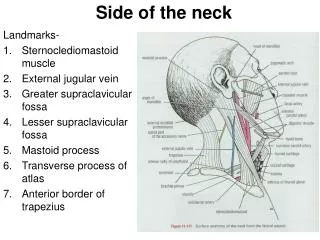
Sternocleidomastoid • Origin: Upper part of manubrium & medial third of clavicle • Insertion: Mastoid process & lateral part of superior nuchal line • Nerve supply: Spinal part of accessory nerve (motor) & ventral rami of C2-3 (proprioceptive) • Action: • Both muscles acting together extend head at atlanto-occipital joint, and flex cervical part of vertebral column • Contraction of one muscle moves the face to the opposite side
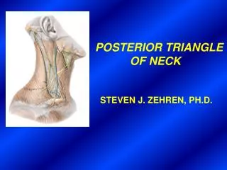
Boundaries • Anterior: Posterior border of sternocleidomastoid • Posterior: Anterior border of trapezius • Inferior: Middle third of clavicle • Roof: skin, superficial fascia, platysma, investing layer of deep fascia • Floor: muscles covered by prevertebral fascia
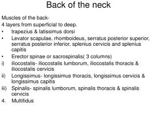
Muscular Floor of the Posterior Triangle From above downward: • Semispinalis capitis • Splenius capitis • Levator scapulae • Scalenus medius • Scalenus anterior may or may not be present
Subdivision of the Posterior Triangle • Subdivided by the inferior belly of omohyoid muscle, into: • Large occipitaltriangle above • Small supraclaviculartriangle below
Omohyoid Muscle • Two bellies: Superior & Inferior joined by intermediate tendon • Attachement: Superior belly to inferior border of hyoid bone, Inferior belly to superior border of scapula & suprascapular ligament • The Intermediate tendon lies deep to sternocleidomastoid, connected to clavicle & the first rib by a loop of deep fascia • Nerve supply:Ansacervicalis (C1,2,3) Superior belly Intermediate tendon Inferior belly Action: Depresses the hyoid bone
Contents • Arteries: • Subclavian (3rd part) • Superficial cervical & suprascapular(branches of thyrocervical trunk, a branch of 1st part of subclavian artery • Occipital, a branch of external carotid artery
Veins: • External jugular vein • Formation • Termination • Tributaries
Nerves: • Branches of cervical plexus • Spinal part of accessory nerve • Brachial plexus
Clinical Notes • Torticollis (wry neck): • Congenital: due to excessive stretching of sternocleidomastoid muscle during labor. • Spasmodic: usually psychogenic • Injury to spinal part of accessory nerve • Injury to brachial plexus • Pleura & Lung injuries in the root of neck • Injury to nerve to platysma