Download
1 / 28
280 likes | 374 Views
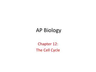
AP Biology. Chapter 12: The Cell Cycle. One cell becoming two. Chromatin vs. Chromosomes appearance within the cell. Fig: 19.4 Coiling up of Chromatin. Somatic cells vs. Germ cells The egg surrounded by sperm. Fig: 12.4 Before and after the S phase.

E N D
AP Biology Chapter 12: The Cell Cycle
Interphase cell (Look at the chromatin in the blue nucleus and the yellow cytoskeleton.)
Chromosome movement LE 12-8b Kinetochore Tubulin subunits Motor protein Microtubule Chromosome
Chromatin condensing Nucleus 10 µm Chromosomes Cell plate Nucleolus LE 12-10 Prometaphase. We now see discrete chromosomes; each consists of two identical sister chromatids. Later in prometaphase, the nuclear envelope will fragment. Prophase. The chromatin is condensing. The nucleolus is beginning to disappear. Although not yet visible in the micrograph, the mitotic spindle is starting to form. Metaphase. The spindle is complete, and the chromosomes, attached to microtubules at their kinetochores, are all at the metaphase plate. Telophase. Daughter nuclei are forming. Meanwhile, cytokinesis has started: The cell plate, which will divide the cytoplasm in two, is growing toward the perimeter of the parent cell. Anaphase. The chromatids of each chromosome have separated, and the daughter chromosomes are moving to the ends of the cell as their kinetochore micro- tubules shorten.
Microscopic view of Mitosis in Onion root tips.Can you identify the stages?
Cell wall Origin of replication Plasma membrane E. coli cell Bacterial chromosome Chromosome replication begins. Soon thereafter, one copy of the origin moves rapidly toward the other end of the cell. Two copies of origin LE 12-11_3 Origin Origin Replication continues. One copy of the origin is now at each end of the cell. Replication finishes. The plasma membrane grows inward, and new cell wall is deposited. Two daughter cells result.
LE 12-15 G0 G1 checkpoint G1 G1 If a cell does not receive a go-ahead signal at the G1 checkpoint, the cell exits the cell cycle and goes into G0, a nondividing state. If a cell receives a go-ahead signal at the G1 checkpoint, the cell continues on in the cell cycle.
G1 S G2 G1 M M M S G2 MPF activity LE 12-16a Cyclin Relative concentration Time Fluctuation of MPF activity and cyclin concentration during the cell cycle
LE 12-16b G1 Cyclin S Cdk M Degraded cyclin G2 accumulation G2 checkpoint Cdk Cyclin is degraded Cyclin MPF Molecular mechanisms that help regulate the cell cycle
Chromosome movement LE 12-8b Kinetochore Tubulin subunits Motor protein Microtubule Chromosome
Cells anchor to dish surface and divide (anchorage dependence). When cells have formed a complete single layer, they stop dividing (density-dependent inhibition). LE 12-18a If some cells are scraped away, the remaining cells divide to fill the gap and then stop (density-dependent inhibition). 25 µm Normal mammalian cells
Cancer cells do not exhibit anchorage dependence or density-dependent inhibition. LE 12-18b 25 µm Cancer cells
Malignant cancer cells from the breast(See the ABNORMAL “crab” shape of the cells.)