Download
1 / 102
1.03k likes | 1.37k Views
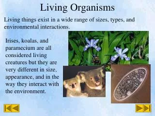
Living Organisms. Living systems are separated from other chemical systems by; . The capacity for replication; The presence of enzymes and other complex molecules; A membrane that separates the internal chemicals from the external chemical environment. Terms applied to cells.

E N D
Living systems are separated from other chemical systems by; • The capacity for replication; • The presence of enzymes and other complex molecules; • A membrane that separates the internal chemicals from the external chemical environment.
Terms applied to cells • Heterotrophs (other-feeder): an organism that obtains its energy from another organism. Animals, fungi, bacteria, and many protistans are heterotrophs. • Autotrophs (self-feeder): an organism that makes its own food, it converts energy from an inorganic source in one of two ways • Photosynthesis is the conversion of sunlight energy into C-C covalent bonds of a carbohydrates. This led to the oxidative metabolism • Chemosynthesis is the capture of energy released by certain inorganic chemical reactions.
Time scale of Evolution • Life emerged at least 3.8 billion years ago. • Simple organic molecules could form and spontaneously polymerize into macromolecules. • No free oxygen but consists CO2 and N2.. Also small amount of H2, H2S and CO. • RNA world-self replicating RNA molecules.
Evolution of cells From the Cell, A Molecular Approach 2nd edition; Cooper; ASM Press & Snauer
4.2 Cell sizes vary with their function • Below is a list of the most common units of length biologists use (metric) Table 4.2
Cell size and shape relate to function Figure 4.2
Why cell size vary? • Smallest cells: • Mycoplasmas; they have the smallest genome • Bulkiest cells: • Bird eggs, young need a lot of food • Longest cells: • Nerve cells, can transmit signals over long ranges
What limits cell size? • Lower limits • What does the cell need to contain? • Must house DNA, proteins, and organelles (in eukaryotes). • Upper limits • It must have enough surface area, why? • Must be able to obtain enough nutrients from the environment.
Prokaryotic Cells • Archaebacteria • Eubacteria • They have plasma membrane • They have nucleoid • They have cytoplasm with ribosomes
Prokaryote (pro=before, karyo=nucleus) From Life: The Science of Biology, 4th Edition Sinauer & WH Freeman
Prokaryotic cells • Very diverse in their metabolic capabilities. • Some archae are found in hot springs • Some of them are photosynthetic. • Some are able to oxidize inorganic ions to obtain energy • prokaryotes are asexual, meaning their offspring nearly always bear the exact characterisics of the parent cell. Division is by binary fission.
Prokaryotic cells • Prokaryotic DNA is organized as a circular chromosome. • DNA is supercoiled • Most of DNA is protein coding
Prokaryotes • In Greek pro means before and karyon refers to nucleus. • Nucleoid(=nucleus like), coiled DNA of a prokaryote. • No organelles in prokaryotes. • Ribosomes (that assemble amino acids) are free in cytoplasm. • Cell membrane surrounds the cell; cell wall protects the cell. In some, there is a sticky coat called a capsule (works like a glu). • Pili and flagella are for attachment and movement.
Procaryote sizes and structures From Molecular Biology of the Cell Third edition; Alberts; Garland
Specialized features of some prokaryotes-1 • Cell wall: Outside the PM. Supports the cell and determines the shape. • It contains peptidoglycan. • It is not a barrier and some toxins can cause disease From Life: The Science of Biology, 4th Edition Sinauer & WH Freeman
Specialized features of some prokaryotes-2 • Capsule: • It encloses cell wall and outer membrane. • It may protect from WBC • It is not necessary for living
Specialized features of some prokaryotes-3 • Mesosome: • It is formed by infolding of the PM • It may aid the movement in & out of the cell of materials. It may also aid the replication of DNA and cell division.
Specialized features of some prokaryotes-4 • Flagella • Bacterium moves with its help • It is anchored to the PM and cell wall
Specialized features of some prokaryotes-5 • Pili • projected from the surface • helps to adhere to another bacteria • shorter than flagella
From the Cell, A Molecular Approach 2nd edition; Cooper; ASM Press & Snauer
Structures of animal cells From the Cell, A Molecular Approach 2nd edition; Cooper; ASM Press & Snauer
Eukaryotic Cells: • Plasma membrane: to define its boundary and retain its content • Membranous subcompartments (organelles): various cellular functions are localized • Nucleus: to house the DNA • Cytoplasm: • Plant cells also have a cell wall outside the PM • Animal cells are usually surrounded by an extracellular matrix.
Membranes in eukaryotic cells • It consists of phospholipids and proteins organized into two layers (Phospholipid bilayer) • It has a polar (hydrophilic) head and two nonpolar (hydrophobic) tails.
Diagram of a phospholipid bilayer From: Life 4th Edition, by Sinauer Associates
MEMBRANE STRUCTURE AND FUNCTION 5.10 Membranes organize the chemical activities of cells • Membranes organize the chemical reactions making up metabolism Cytoplasm Figure 5.10
Biological membranes: • To regulate molecular traffic from one side to another • To restrict the passage of materials, especially polar ones, since its hydrophobicity of its interior. • To allow interactions amongst the cells. (i.e. recognition of WBC). • To provide energy (mitochondria and choloroplast)
5.11 Membrane phospholipids form a bilayer • Phospholipids are the main structural components of membranes • They each have a hydrophilic head and two hydrophobic tails Head Symbol Tails Figure 5.11A
In water, phospholipids form a stable bilayer • The heads face outward and the tails face inward Water Hydrophilicheads Hydrophobictails Water Figure 5.11B
The plasma membrane of an animal cell Glycoprotein Carbohydrate (of glycoprotein) Fibers of the extracellular matrix Glycolipid Phospholipid Cholesterol Microfilaments of the cytoskeleton Proteins CYTOPLASM Figure 5.12
Biological membranes: From http://www.biosci.uga.edu/almanac/bio_103/notes/may_15.html.
Structure of an animal cell Fromhttp://www.biosci.uga.edu/almanac/bio_103/notes/may_15.html.
Nucleus • Nuclear envelope: Inner and outer nuclear membranes • Nuclear pores • Nucleolus From: Life 4th Edition, by Sinauer Associates
Liver Cell Nucleus From: www.DennisKunkel.com
Nuclear envelope and nuclear pores From: Life 4th Edition, by Sinauer Associates From: www.DennisKunkel.com
Nucleus • Chromatin: DNA associated with proteins, forms long fibers. • Each fiber constitutes a chromosome. • Chromosomes condense during mitosis/meiosis. • Chromosomes are enclosed within a nuclear envelope, a double membrane with pores. • Nucleolus consists of parts of the chromatin DNA combined with RNA and proteins (components of ribosomes are made).
Cytoplasm • Organelles • cytoskeleton: maintain the shape of the cell as well as anchoring organelles, moving the cell and controlling internal movement of structures • Microtubules • Actin • Intermediate filaments
Many cell organelles are related through the endomembrane system • The endomembrane system is a collection of membranous organelles • These organelles manufacture and distribute cell products • The endomembrane system divides the cell into compartments • Endoplasmic reticulum (ER) is part of the endomembrane system
Endomembrane System • Contains • Rough ER (makes membrane and proteins) • Smooth ER (makes lipids, destroys toxins, stores calcium • Golgi • Lysosomes • Vacuoles • Nuclear envelope
Rough ER • Contains ribosomes. • It makes membrane when necessary. • Some proteins made by RE are inserted into the ER membrane. • Phospholipids are made by ER enzymes. • ER membrane enlarges. • Makes proteins secreted by the cell. • Secretory proteins, e.g., antibody, a defensive molecule. Ribosomes synthesize the proteins of the antibody, they are assembled in the ER. Short chains of sugars are linked (glycoprotein), are transported in the transport vesicle, that buds off.
Transport vesiclebuds off 4 Ribosome Secretory(glyco-) proteininside transportvesicle Sugarchain 3 Glycoprotein 1 2 ROUGH ER Polypeptide 4.8 Rough endoplasmic reticulum makes membrane and proteins • The rough ER manufactures membranes • Ribosomes on its surface produce proteins Figure 4.8
Ribosomes From: Life 4th Edition, by Sinauer Associates From: www.DennisKunkel.com
Smooth ER • Continuous with RE, and lack ribosomes. • It has enzymes within the membrane. • Synthesize lipids (fatty acids, phospholipids, steroids) depending on the type of the cell. • Regulate the amount of sugar released from liver cells into the bloodstream. • Other enzymes break drugs, detoxify. • SER increase by exposure to drugs and produce tolerance. Sometimes it can not distinguish between drugs, so tolerance to a wide range of drugs occurs. (Barbiturate, a sedative, may decrease the effectiveness of antibiotics.
4.9 Smooth endoplasmic reticulum has a variety of functions • Smooth ER synthesizes lipids • In some cells, it regulates carbohydrate metabolism and breaks down toxins and drugs
SMOOTH ER ROUGHER Nuclearenvelope Ribosomes SMOOTH ER ROUGH ER Figure 4.9
Endoplasmic Reticulum From: Life 4th Edition, by Sinauer Associates From: www.DennisKunkel.com
4.10 The Golgi apparatus finishes, sorts, and ships cell products • The Golgi apparatus consists of stacks of membranous sacs • These receive and modify ER products, then send them on to other organelles or to the cell membrane
Golgi Apparatus • Flattened sacs looking like a stack of pitabread. • Sacs are not interconnected. • A cell may contain a few or a lot of them, depending on its activity. • It serves as a molecular warehouse and finishing factory through modification of substances manufactured by ER.