Download
1 / 26
260 likes | 365 Views
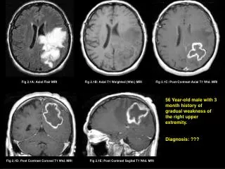
Rohit Gautam, Jayanthi Sivaswamy CVIT, IIIT Hyderabad, Hyderabad, India Ravi Varma KIMS Hospital, Hyderabad, India. A method for motion Detection and Categorization in perfusion weighted MRI. Introduction. MRI.

E N D
Rohit Gautam, Jayanthi Sivaswamy CVIT, IIIT Hyderabad, Hyderabad, India Ravi Varma KIMS Hospital, Hyderabad, India A method for motion Detection and Categorization in perfusion weighted MRI
Introduction MRI • MRIfor brain - Application of nuclear magnetic resonance (NMR) to create images of human brain. From MRI to Perfusion MRI • Many neurological disorders can be detected using abnormal blood flow. • Perfusion MRI utilizes this blood flow information in disease diagnosis. PerfusionMRI
What is Perfusion MRI ? • Perfusion is the delivery of oxygen and nutrients to the cells via capillaries. • A bolusinjected into patient’s blood is tracked over time. • It provides information regarding rate of blood flow, which helps to determine the affected regions in brain on the onset of disorder. • Acquired data is a 3D time-series. Volume nwout After Bolus wash-out Before Bolus wash-in Bolus in transit nwin 1 N Time-points
Problem Patient motion during MRI scan (> 2 minutes) misaligns (corrupts) the acquired data. Aim - Motion detection and categorization of volumes corrupted due to patient motion. Difficulties • Simultaneous local (non-uniform variation in image contrast due to bolus) and global (motion) changes. • Current scenario: Motion correction is a time-limiting step in PWI analysis [ Straka et al. JMRI 07].
1 Before bolus wash-in Motion No variation in intensity Bolus in transit Motion Non-uniform Variation in intensity After bolus wash-out Motion N No variation in intensity
Why motion correction ? Perfusion parameters obtained from motion corrupted data vary with degree of motion. Error in CBV estimation Error in TTP estimation TTP: Time to Peak CBV: Cerebral Blood Volume
Division of Time-Series • The signal intensity in perfusion MRI varies proportionally with bolus concentration. • A gamma-variate function (GVF) can model the change in concentration of bolus with time. • We fit GVF on the mean-intensity perfusion curve µa(n) to estimate GVF-fit mean intensity curve µg(n). • Using µg(n), we divide the time-series into 3 sets: Set1: pre-wash-in, Set2: transit and Set3: post-washout sets Wash-in Time point Wash-out Time point
Intensity Correction • Bolus is present only in Set2. Hence, intensity correction is required for bolus affected regions in these volumes before motion detection. • A volume (F) is segmented into normal (Fnormal) and bolus affected (Fbolus) regions using clustering technique. • F is then intensity corrected: where, µg(n) is the GVF-fit-mean intensity curve.
Intensity Correction Absolute Difference Slice 1 Slice 2 Reduction in absolute intensity difference Intensity Correction Ideally, these should be 0 Intensity Corrected Slice 2 Absolute Difference Slice 1
Motion Detection For each mean shifted blocks of fixed size around each pixel, the flow vector is given by: where S is the cross power spectrum. Block wise Phase Correlation Extract Central Slices Block wise Phase Correlation Process is accelerated by down-sampling of central slices.
Motion Flow Maps Slice 1 Slice 2 Slice 1 Slice 1 Slice 2 Slice 2 Un Un Un Vn Vn Vn Bolus present and No motion Bolus absent and Minimal motion Bolus absent and Mild motion
Net Entropy Metric • For a given time series {Fn; n=1…N} , the net entropy (Hn) of flow fields (Un;Vn)is given by: • The net entropy is 0 for no motion and increases with degree of motion.
Net Entropy Profile 1 Zero net entropy even in the presence of bolus. 5 8 40 33 Wash-in Time-point Wash-in Time-point
Does Intensity Correction help ? A non-zero net entropy even in the absence of motion
Time Analysis of motion detection Large reduction in computation time
Motion Categorization • Peak entropy of flow fields is used to quantify the degree of motion. • The peak entropy Hpeakof the flow fields is found by: where, Hnis the net entropy.
Such a small motion cannot be detected. Peak entropy can distinguish between different motion categories. Entropy values for different motion categories for image size – 32x32 and block size 8x8
Comparison of our approach • We compare the efficiency of motion detection method by applying it prior to existing motion correction algorithms. [1] Kosior et al., JMRI 2007. [2] Straka et al., JMRI 2010. [3] Tanner et al., MICCAI 2000.
Conclusions • We proposed a motion detection method that is immune to intensity changes due to injected contrast agent. • We achieved a large reduction in time (~37%) required for motion correction by rejecting the stationary volumes. • The detection method can be made to be fast but the sensitivity to minimal motion maybe compromised.