Download
1 / 46
470 likes | 635 Views
Chapter 7, Membranes. Cell and Molecular Biology. Cellular Membranes. Membranes were “predicted” to surround cells long before membranes were seen Membranes were predicted to be mostly lipid based on the diffusion of lipid soluble molecules into cells

E N D
Chapter 7, Membranes Cell and Molecular Biology
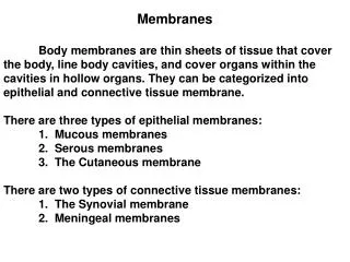
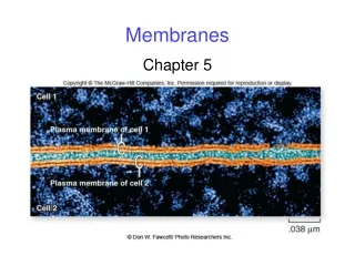
Cellular Membranes • Membranes were “predicted” to surround cells long before membranes were seen • Membranes were predicted to be mostly lipid based on the diffusion of lipid soluble molecules into cells • Other properties of membranes suggested they also contained protein • In the 1950’s, the EM allowed visualization of membranes
Cellular Membranes • In addition to the plasma membrane, which separates the cell’s interior from the external environment, the ER, nucleus, mitochondria, chloroplasts, lysosomes, peroxisomes, and transport vesicles are all surrounded by membrane • The membrane isolates various “compartments” within the cell • Many processes in the cell occur either on, in, or in association with membrane surfaces
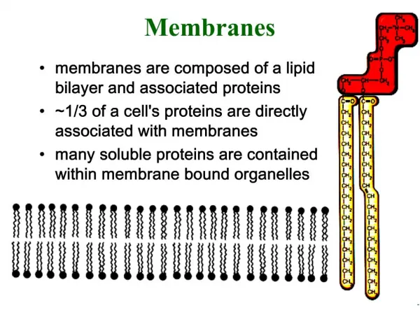
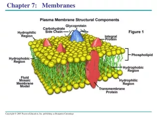
Fluid Mosaic Model of Membrane Structure • The current understanding of membrane structure and composition has led to the formation of the “fluid mosaic model” • In this model, the membrane is seen as a fluid lipid bilayer (composed mostly of phospholipids), with a mosaic of various proteins “floating” on and in the lipid layer • Some membranes, such as the mitochondrial inner membrane, contain more protein than lipid
Key Features of the Fluid Mosaic Model • 1) Membranes are arranged in the form of a lipid bilayer, which is interrupted by embedded proteins • 2) The lipid bilayer is fluid • 3) There are several ways in which proteins interact with the membrane • A) Integral membrane proteins • B) Peripheral membrane proteins • C) Lipid anchored proteins
Membrane Proteins • Integral membrane proteins: proteins which are embedded within the lipid bilayer: these proteins are held in the membrane by hydrophobic forces • Peripheral membrane proteins: these proteins are attached to the surface of the membrane, and can be on either the outer or inner face of the membrane. These are usually held in place by ionic forces with the phospholipid head groups or other proteins • Lipid anchored proteins: these proteins are outside the layer of the membrane, but are covalently attached to lipids which are within the membrane
Different Membranes Contain Different Amounts of Proteins and Different Lipid Compositions
Lipids of Membranes • Most lipids in membranes are phospholipids, glycolipids, and steroids • The exact composition of a membrane varies with the cell, the organism, and the organelle • Membrane lipids are amphipathic, that is, they have a polar or hydrophilic end (the head group) and a hydrophobic (lipid) portion
Membrane Phospholipids • Numerous phospholipids are found in membranes • All have a phosphate group covalently attached to the hydrophobic lipid Ended here monday
Membrane Glycolipids • Numerous glycolipids are found in membranes • These have a carbohydrate covalently attached to the lipid • Steroids (cholesterol) represent a third important class of membrane lipids
Different Membranes have Different Phospholipid Compositions Different membranes in a cell, or in different cell types, have different lipid compositions, giving the membranes different properties.
Membrane Transition Temperature • Altering the lipid type in a membrane changes the temperature at which the membrane changes from a gel to a fluid • Cell membranes are generally fluid at physiological temperatures • Cells maintain a fluid membrane by changing the lipids in the membrane
Transition Temperatures • Long chain fatty acids: higher transition temps (more solid) • Unsaturated fatty acids: lower transition temps (more liquid) • Steroids: inflexible rings: more solid • But Steroids prevent “packing” of fatty acid chains, prevent gelling, more liquid • Steroids (cholesterol) act as a structural “buffer” to prevent changes in the gel state of membranes
Transition Temperatures • Organisms (Plants and Microbes) adjust to lower temperatures by increasing the degree of lipid unsaturation or decreasing average fatty acid chain length • This allows these organisms to maintain membrane fluidity • In animals, fatty acid composition of membranes is influenced by diet • Changing your diet can alter the membrane makeup
Lipid Movement Within a Membrane • Lipids are relatively unconstrained in lateral diffusion • Transverse diffusion (flip flop) occurs very slowly • In other words, lipids can move around on the membrane, but don’t usually move from one side to the other (from one leaflet to the other) • Proteins called phospholipid translocators can speed the movement of phospholipids from one side of the membrane to the other
Techniques for Studying Membrane Composition • Red Blood Cells (RBCs) can be broken open and membrane vesicles prepared. • If Mg2+ ions are included in the buffer, the membranes retain their normal orientation. • If no Mg2+ is included, the vesicles are “inside out” : the side of the membrane which normally faces the inside of the RBC facing out.
Determining the Composition of Inner and Outer Membrane Leaflets • Isolate either “right side out” or “inside out” membrane vesicles • Treat the vesicle with phospholipase, which • A) digests phospholipids • B) cannot pass through the membrane • Determine which lipids were degraded • Phosphatidylcholine and sphingomyelin are mostly in the outer leaflet • Phosphatidylethanolamine and phosphatidylserine are mostly in the inner leaflet
“Inner” and “Outer” Leaflets • Since transverse diffusion is limited, the inner and outer leaflets of a membrane may have very different compositions • They may vary in the type of phospholipids present • They may vary in the saturation level of the fatty acids (less saturated outer membrane, less fluid) • Glycolipids are seen only in the outer leaflet of the plasma membrane
Function of Membrane Proteins • Four major functions of membrane proteins • Transport: move molecules across the membrane • Receptors: carry signals across the membrane • Attachment: provide structure and form • Metabolism: enzymes
Labeling of Proteins on a Single Surface of Membranes • Selective labeling of proteins allows you to determine the orientation of the protein in the membrane
Major Types of Membrane Proteins • Membrane proteins are characterized by the ways in which they interact with the membrane
Major Types of Membrane Proteins • Peripheral membrane proteins interact with only one side of the membrane (ionic interactions), and are easily removed by high salt buffers • Integral membrane proteins usually span the membrane (can be labeled from either side) and require strong detergent (SDS) extraction to be removed • Integral membrane proteins are described by the number of “passes” through the membrane, and often contain multiple subunits
Hydropathy Plot • Plots of the hydrophobic/hydrophylic nature of a protein as you “scan” along the protein’s sequence are called hydropathy plots • Hydropathy plots are used to predict regions of a protein which will be within a membrane
Movement of Proteins Within a Membrane • Various techniques have been used to examine the ability of proteins to move within a membrane • Some are found to be unconstrained in their diffusion
Movement of Proteins Within a Membrane • Photobleaching is a technique to see the diffusion of a single type of protein (labeled with a fluorescent marker)
Protein Movement • Some proteins are relatively free to move • Many membrane proteins are constrained in their ability to move • “Domains” exist within membranes: proteins may be free to move, but only within a certain region of the membrane • Motion is limited by several mechanisms • Aggregation • anchoring to the cytoskeleton or extracellular matrix • barriers such as tight junctions
RBC Proteins • The Red Blood Cell (RBC) has been a good model for studying membrane proteins because it contains few proteins • SDS PAGE shows a relatively simple protein band pattern Ended here wed
Techniques for Studying Membrane Composition • Red Blood Cells (RBCs) can be broken open and membrane vesicles prepared • If Mg2+ ions are included in the buffer, the membranes retain their normal orientation • If no Mg2+ is included, the vesicles are “inside out” : the side of the membrane which normally faces the inside of the RBC facing out
Major Types of Membrane Proteins • Membrane proteins are characterized by the ways in which they interact with the membrane
RBC Proteins • Six major proteins in RBC membrane • Extraction with high ionic strength solutions removed spectrin, ankyrin, band 4.1, actin (what does this tell you?) • These 4 proteins labeled only in inside out vesicles (what does that tell you?) • Glycophorin and band 3 are only removed after SDS extraction (what does this tell you?) • “Periodic Acid- Schiff reagent” (PAS), which reacts with (labels) carbohydrates, labels glycophorin and band 3
Quiz 6 • 1. A. What are the three types of lipids which make up most of the membrane? • B. What is the distinguishing characteristic of each type? • 2. A. What are the three types of proteins associated with membranes? • B. What is the distinguishing characteristic of each type?
RBC Integral Membrane Proteins • Proteins on “inside out” or “right side out” vesicles were labeled with radioactive molecules • Glycophorin and “Band 3” labeled in both types of vesicles (what does that mean?) • Protein isolated after labeling was digested (with a protease) and the fragments separated by electrophoresis • Different fragments were labeled in the two types of vesicles (what does that mean?)
Glycoproteins of RBC • Treatment of right side out vesicles with protease released carbohydrates into medium • Similar treatment of inside out vesicles did not release carbohydrate (what does this mean?) • Carbohydrates could be enzymatically labeled in right side out but not inside out vesicles • Carbohydrates on proteins face the non- cytoplasmic side of the membrane (the outside of the plasma membrane, or the organelle interior of other membranes)
Glycoproteins of RBC • Labeling and digestion experiments have determined the arrangement of glycophorin and “band 3” glycoproteins
Glycoproteins of RBC • Glycophorin turns out to be a single pass protein, 113 amino acids, with 16 carbohydrate groups attached • The N-terminal region (amino terminus) faces away from the cytoplasm: this is where the carbohydrates are attached • The C-terminal region (carboxy terminus) faces the cell interior: this is mostly hydrophilic amino acids • A hydrophobic alpha-helix spans the membrane
Glycoproteins of RBC • Band 3 proteins is a dimer: two identical polypeptide chains ( a homodimer) • Both the N-terminus and the C-terminus of each subunit face the cytoplasmic side • Multiple (about 12) hydrophobic alpha-helical segments span the membrane • A single hydrophilic carbohydrate chain is attached to the protein, on the non-cytoplasmic side
N-Linked and O-Linked Glycosylation • There are two different processing events which attach carbohydrates to proteins: • N-linked glycosylation attaches carbohydrate to the nitrogen of asparigine residues. This occurs in the ER. • O-linked glycosylation attaches carbohydrates to the oxygen of serine or threonine. This process occurs in the Golgi.
Glycosylation on Modified Amino Acids • The amino acids (residues) proline and lysine are sometimes modified to contain a hydroxyl group. This occurs in the ER. • These modified residues can then be glycosylated (O-linked)(in the Golgi).
Common Sugar Groups in Glycoproteins • Galactose, Mannose, N-acetylglucosamine, and sialic acid are the most common carbohydrate monomers in the glycosylated proteins.
Oligosaccharides on Glycoproteins are often in Complex, Branched Arrangements Note that Sialic Acid is negatively charged.
RBC Peripheral Membrane Proteins • Spectrin ( an intermediate filament protein) and actin (actin microfilaments) are cyto- skeleton proteins • Band 4.1 links glycophorin to the cytoskeleton • Ankyrin links (anchors) band 3 protein to the cytoskeleton • The interactions between these proteins appear to give the RBC much of its characteristic “bi-concave” shape
RBC Protein Arrangement • “Cell and Molecular Biology” is an attempt to elucidate the way in which cells work.
Some Proteins are Complex Structures • The photosynthetic reaction center of Rhodopseudomonas vividis contains 4 different subunits: two are multipass integral membrane peptides, one is an integral protein facing the cytosol, and one is a peripheral peptide facing the exterior of the cell