Download
1 / 47
480 likes | 636 Views
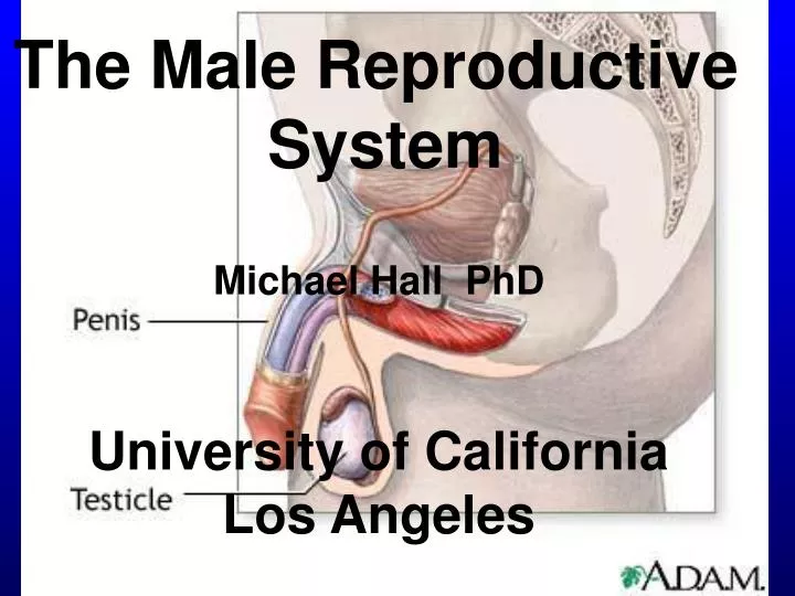
The Male Reproductive System. Michael Hall PhD. University of California Los Angeles. Components of the Male Reproductive System. Embryology of Gonad Development. Mesenchyme. Genital ridge. 6 weeks (male = female). Medulla Cortex. 7-8 weeks. Female. Male. Functions of Testes.

E N D
The Male Reproductive System Michael Hall PhD University of California Los Angeles
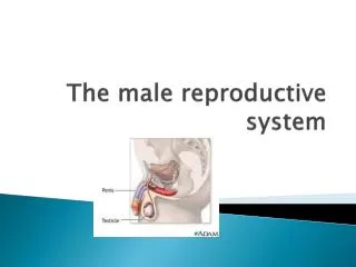
Components of the Male Reproductive System
Embryology of Gonad Development Mesenchyme Genital ridge 6 weeks (male = female) Medulla Cortex 7-8 weeks Female Male
Functions of Testes Spermatogenesis-production of sperm a. spermatocytogenesis-mitotic division of spermatogonia b. meiosis-production of haploid spermatids c. spermiogenesis-maturation of spermatids to spermatozoa 2. Steroidogenesis-production of steroid hormones
Testis Head of epididymis Vas deferens Efferent ducts Seminiferous tubule Body of epididymis Septum Mediastinum testis Tunica albuginea Rete testis Tunica vaginalis Tubulirecti Tail of epididymis
T. albuginea Seminiferous tubules
Components of the seminiferous epithelium Lumen (DNA) Spermatozoa (N) Spermatid (N) Secondary spermatocyte (2N) Sertoli cell cytoplasm Primary spermatocytes Sertoli cell nucleus (4N) Spermatogonium (2N) Basal lamina CT
Spermatogenesis-production of sperm a. spermatocytogenesis-mitotic division of spermatogonia (2N) to primary spermatocytes (4N) b. meiosis-two successive divisions producing haploid spermatids (N) c. spermiogenesis-maturation of spermatids to spermatozoa (non-motile)
Sertoli Cells Lumen (DNA) Spermatozoa (N) Spermatid (N) Secondary spermatocyte (2N) Sertoli cell cytoplasm Primary spermatocytes Sertoli cell nucleus (4N) Spermatogonium (2N) Basal lamina CT
Sertoli cells form tight junctions Junctional complex JC
Functions of Sertoli Cells Support, protection, and nutritional regulation of the developing spermatozoa 2. Phagocytosis of residual bodies 3. Secretion of testicular fluid into lumen 4. Production of Androgen Binding Protein (ABP) 5. Secretion of Inhibin (suppresses FSH secretion)
Components of the seminiferous epithelium Lumen Spermatozoa (N) Spermatid (N) Secondary spermatocyte Sertoli cell cytoplasm (2N) Primary spermatocyte Sertoli cell nucleus (4N) Spermatogonium (2N) Basal lamina
Sertoli cells in seminiferous tubule Sertoli cells Sertoli cell
Leydig cells Leydig cells Leydig cells lie in the CT between the seminiferous tubules Early spermatids
Leydig cells Seminiferous tubule Seminiferous tubule Leydig cells Seminiferous tubule Seminiferous tubule
Function of Leydig cells and Testosterone The prime function of the Leydig cells is the production of testosterone. Testosterone has many separate but interrelated functions: a). differentiation and maturation of sperm in the seminiferous tubule (200x testosterone) b). Development and secretory activity of accessory sex glands ( seminal vesicle, prostate and bulbourethral glands c). development of male secondary sex characteristics (facial and pubic hair, muscularity, libido, obnoxious behavior, voice etc.)
Hormonal control of spermatogenesis Hypothalamus GnRH 1.LH stimulates synthesis of male sex hormones by Leydig cells LH 1 2.FSH stimulates Sertoli cells to synthesize ABP FSH 2 Inhibin from Sertoli cells 3.Leydig cells produce testosterone into blood into tubule 3 Seminiferous tubule
Components of the Genital Duct System seminal vesicle prostate gland urethra b-u gland efferent ducts epididymis vas deferens rete testis
Components of the Genital Duct System seminal vesicle prostate gland ampulla urethra b-u gland efferent ducts epididymis vas deferens rete testis
Efferent duct Epididymis smoothmuscle sc cilia smoothmuscle
Components of the Genital Duct System seminal vesicle prostate gland ampulla urethra b-u gland efferent ducts epididymis vas deferens rete testis
Efferent duct Epididymis smoothmuscle sc cilia smoothmuscle
Components of the Genital Duct System seminal vesicle prostate gland ampulla urethra b-u gland efferent ducts epididymis vas deferens rete testis
Vas deferens IL Smooth muscle layers C OL
Vas deferens stereocilia smooth muscle
Components of the Genital Duct System seminal vesicle prostate gland ampulla urethra b-u gland efferent ducts spermatic cord epididymis vas deferens rete testis
Spermatic cord vas deferens pampiniform plexus cremaster muscle
Pampiniform plexus of spermatic cord Nerve Arteries and veins
The Accessory Sex Glands prostate gland seminal vesicle urethra b-u gland
Seminal vesicle Cross section of a single, highly coiled seminal vesicle surrounded by smooth muscle SM 1 2. High magnification of the mucosa of a seminal vesicle Epithelium 2
Prostate Gland Bladder Urethra Smooth muscle Capsule Mucosal glands Submucosal glands Main glands
Prostate Gland Glands and ducts SM Prostatic concretions
Prostate Gland Glands and ducts PC Smooth muscle and elastic fibers Epithelium
Bulbourethral gland prostate gland seminal vesicle ampulla urethra b-u gland efferent ducts epididymis vas deferens rete testis
The Penis- has two functions: Urination Reproduction
The Penis erectile tissue Corpora cavernosa Tunica albuginea Skin Corpus spongiosum Urethra
Penis: Erectile Tissue elastic fibers and smooth muscle endothelial cells line cavernous spaces trabeculae
The Penis Blood flow to corpus cavernosa and mechanism of erection Helicine artery
Mechanism of action of Viagra Penile vascular smooth muscle cell relaxation GTP Viagra (Loss of erection)
See, the problem is that God gives men a brain and a penis and only enough blood to run one of them at a time. Robin Williams
Semen Semen consists of spermatozoa and secretions of the accessory sex glands, which follow one another in a definite sequence: During arousal: Bulbourethral gland secretes mucous-like secretion which lubricates the urethra At ejaculation: 1. Prostate gland secretion—proteolytic enzyme rich 2. Spermatazoa are ejected 3. Seminal vesicle secretion follows—nutrient rich NB: Spermatozoa comprise only 5% of semen
Thank you It’s been great—but it’s time to be going home (my wife’s wondering where I’ve gone!) bon voyage et bonne chance!