Download
1 / 35
530 likes | 1.46k Views
DEVELOPMENT OF ARTERIES AND VEINS. AORTIC ARCHES. Arch Arteries . Placental Vein Liver Heart Truncus Arteriosus Each Arch artery Dorsal aorta Aortic Sac. Arch Arteries Arch 1 mainly lost, form part of maxillary artery Arch 2 stapedial arteries Arch 3

E N D
Arch Arteries Placental Vein Liver Heart TruncusArteriosus EachArchartery Dorsal aorta Aortic Sac • Arch Arteries • Arch 1 • mainly lost, form part of maxillary artery • Arch 2 • stapedial arteries • Arch 3 • common carotid arteries • internal carotid arteries • Arch 4 • left forms part of aortic arch • right forms part right subclavian artery • Arch 6 • left- part of left pulmonary artery • right- part of right pulmonary artery
DEVELOPMENT of the AORTIC ARCHES • The first pair of aortic arches is formed by the curving of the ventral aorta to meet the dorsal aorta, these will eventually contribute to the external carotid arteries. • The second pair of aortic arch arteries appear in week four. • These regress rapidly and only portion is left which forms the stapedial and hyoid arteries. • The third pair of the aortic arch arteries appear around the end of the fourth week. • Thesewill give rise to the common carotid arteries and proximal portion of the internal carotid arteries. • The distal portion of the internal carotid arteries are formed by the cranial portions of the dorsal aorta.
DEVELOPMENT of the AORTIC ARCHES • The fourth aortic arch arteries develop soon after the third arch arteries. • Their development differ on the left from that on the right. • On the left side, it persists, connecting the ventral aorta to the dorsal aorta, forming the aortic arch. • On the right it forms the proximal portion of the right subclavian artery. • The fifth pair of aortic arch arteries are rudimentary and do not develop into any known vessels, this pair of aortic arch arteries are not seen in many embryo specimens. • The sixth aortic arch arteries develop in the middle of the fifth week. • The proximal portions develop into the right and left pulmonary arteries, • The distal portion of the left aortic arch artery develop into the ductus arteriosus.
ARTERIEL SYSTEM DERIVATIVES • Aortic sac (R and L sides of sac): brachiocephalic artery (right) and part of the ascending aorta (left), large parts of common carotid arteries • 1st aortic arch (R and L): part of the maxillary artery and external carotid arteries • 2nd aortic arch (R and L): part of the hyoid and stapedial arteries, part of external carotids • 3rd aortic arch (R and L): part of common carotids and first part of the internal carotids • 4th aortic arch (R): small proximal part of the right subclavian artery • 4th aortic arch (L): small part of arch of the aorta just proximal to the left subclavian artery • 6th aortic arch (R): proximal part of right pulmonary artery • 6th aortic arch (L=ductus): proximal left pulmonary artery and and ligamentum arteriosum • Ductus arteriosus:ligamentum arteriosum • Dorsal aorta (R and L): part of right subclavian, descending aorta below left subclavian • Unpaired ventral (or vitelline) arteries: celiac, superior mesenteric, and inferior mesenteric arteries • Paired dorsal segmental arteries: intercostal arteries and vertebral arteries • Umbilical arteries: internal iliac, superior vesical arteries, medial umbilical ligaments
DEVELOPMENT of the SYSTEMIC VEINS • At day 21 the there is a common atrium as a result of fusion of the two endocardial tubes with two sinus horns, a left and a right horn, representing the unfuses ends of the endocardial tubes [Colvin 1998]. These two horns will form the sinus venosus. • As the bulboventricular portion of the ventricle grows and bends it will form the bulboventricular bend. • This will result in a ventrally situated ventricular chamber(s), and a dorsally situated atria. The sinus venosus lie dorsal to the atria. • The following veins drain into the sinus venosus on each side: • The common cardinal vein, which drains from • the anterior cardinal vein (draining the cranial part of the embryo) • and the posterior cardinal vein (draining the caudal part of the embryo), • the umbilical vein (connecting the heart to the primitive placenta) • and the vitelline vein(draining the yolk sac, GI system and the portal circulation). At 4 weeks gestation three systemic veins connect to each of the two sinus horns
DEVELOPMENT of the SYSTEMIC VEINS • At week four the sinus venosus opens to the common atrium. • In week 7, the sinoatrial communication become more right wards, to connect to the right atrium. • At 8 weeks the distal end of the left cardinal vein degenerates, • The more proximal portion of left cardinal vein will now connect through the anastomosing vein (left brachiocephalic vein) to the right anterior cardinal vein (right brachiocephalic vein), thus forming the superior vena cava. • The left posterior cardinal vein also degenerates, • The left sinus horn receiving venous drain from the heart become the coronary sinus. • The right vitelline vein become the inferior vena cava, • Theright posterior cardinal vein become the azygos vein. • All this is completed in the eighth week of gestation. At 7 weeks gestation the left vitteline and umbilical veins as well as the right umbilical veins degenerate.
DEVELOPMENT of the SYSTEMIC VEINS • The left umbilical veindegenerates • and the right umbilical veinconnects to the vitelline system through the ductus venosus (which is derived from the vitelline veins). • If the anterior cardinal vein does not degenerate on the left side, as it normally does, the future brachiocephalic vein will continue to drain into the sinus venosus, • This will become known as the left superior vena cava and it would drain to the coronary sinus At 8 weeks gestation, the portion closer to the heart of the left anterior cardinal vein degenerates and the anterior cardinal vein connects to the right sided one through a bridging vein
VENOUS SYSTEM DERIVATIVES • Ductus venosus:ligamentum venosum • Umbilical vein (L): round ligament (ligamentum teres) • Vitelline vein (R): superior mesenteric vein and the inferior vena cava • Vitelline vein (L): most of the portal vein • Anterior cardinal veins (R and L): internal jugular veins (left brachiocephalic vein is an anastomosis) • Anterior cardinal vein (R): part of superior vena cava and right brachiocephalic vein • Common cardinal vein (R): proximal part of superior vena cava • Common cardinal vein (L): lateral part of coronary sinus and oblique vein of left atrium • Posterior cardinal vein (R): part of azygos vein and common iliac veins • Supracardinal veins: hemiazygos vein (L) and caudal part of azygos vein (R) • Subcardinal vein (R): renal segment of inferior vena cava
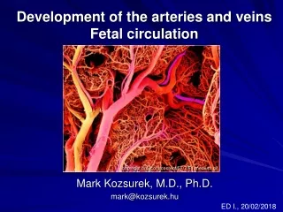
FETAL CIRCULATION • The fetal circulation functions to supply nutrients and oxygen and remove excretory products via the placenta. The vessels supplying oxygenated blood from the placenta are the placental veins. Those returning deoxygenated blood are the placental arteries. • The normal circuit is considered in the submenu in terms of the passage of blood from the: • placenta through the liver • periphery through the heart • The presence of three vessels which bypass longer routes or alternative vascular beds ensures that • the first branches of the aorta receive relatively well-oxygenated blood. • These shunts are the ductus venosus of the liver • and the foramen ovale and ductus arteriosus of the heart.
CIRCULATION CHANGES at BIRTH • The key events underlying changes to the fetal circulation after birth are the: • initiation of respiration • ending of placental blood flow • The circulation changes at birth may be considered in terms of changes to the: • umbilical arteries • umbilical veins • ductus venosus • ductus arteriosus • foramen ovale