Download
1 / 29
380 likes | 1.03k Views
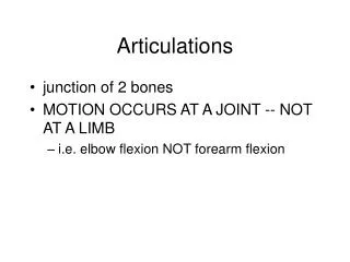
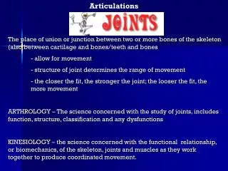
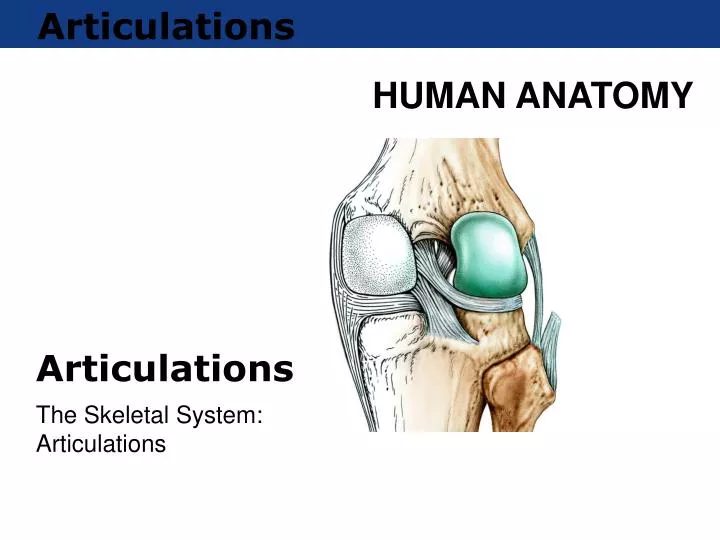
Articulations. Articulations. The Skeletal System: Articulations. Introduction. Joints , or articulations, are connections between bones that may or may not permit movement. Cartilage, fluid, or dense connective tissues is usually involved in holding joints together.

E N D
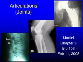
Articulations Articulations The Skeletal System: Articulations
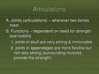
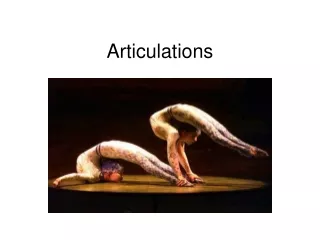
Introduction • Joints, or articulations, are connections between bones that may or may not permit movement. • Cartilage, fluid, or dense connective tissues is usually involved in holding joints together. • Joints maybe classified structurally or functionally
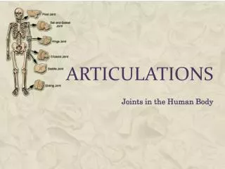
Classification of Articulations - Structural Structural Classification 1) Fibrous – No joint cavity – fibrous tissue a) Suture – very thin fibrous tissue if fused – synostoses b) Syndesmosis – broader fibrous tissue fontanel c) Gomphosis – peg in cone example: tooth in jaw 2) Cartilaginous – No joint cavity – Cartilage a) Synchondrosis – hyaline cartilage epiphyseal plate b) Symphysis – fibrocartilage symphysis pubis and intervertebral disks 3) Synovial – Joint (Synovial) cavity a) Gliding – intertarsal b) Hinge – knee and ankle c) Pivot – atlantoaxial d) Condyloid or Ellipsoidal – wrist e) Saddle – thumb f) Ball and Socket – should and hip
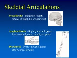
Classification of Articulations Functional Functional Classification of Joint 1) Synarthroses – Immovable Joints a) Suture b) Gompohsis c) Synchondrosis 2) Amphiarthrosis – Slightly movable joints a) Symphysis – pubic symphysis b) Syndesmosis - tibia and fibula 3) Diarthrosis – Freely movable a) Gliding – gliding b) Hinge – flex and extend (1 plane) c) Pivot – rotation – 1 plane d) Condyloid or Ellipsoidal – 2 planes flex, extend, abduct and adduct e) Saddle – – 2 planes flex, extend, abduct and adduct f) Ball and Socket – (3 planes) flex, extend; abduct adduct; and medial and lateral rotation
Synarthroses (Immovable Joints) • Sutures are joints found only in the skull. • Bony edges interlock and short dense connective tissue fiber hold the bones together. • A gomphosis is the joint between a tooth and the alveolar fossa of the maxillae or mandible. • Periodontal ligaments (PDL) hold the tooth to the bone in the gomphosis. • A synchondrosis is a joint in which hyaline cartilage separates the ends of the bones involved in the joint. • A synostosis occurs if bones fuse together to form one bone.
Amphiarthroses (Slightly Movable Joints) • A syndesmosis occurs when to bones are connected by relatively long connective tissue ligaments. • Connecting bones using a fibrocartilage pad forms a symphysis.
Diarthroses (Freely Movable Joints) • Synovial joints are typically found at the ends of long bones in the upper and lower limbs. • All synovial joints have six basic characteristics: • A joint capsule • Articular cartilages • A joint cavity filled with synovial fluid • A synovial membrane lining the joint capsule
Synovial Joints Figure 8.1 Structure of a Synovial Joint
Synovial Fluid • Synovial fluid has three functions: • Lubricates the surfaces of the articular cartilages on the ends of the bones. • Nourishes the chondrocytes by entering and exiting the articular cartilages due to the forces acting on the joint. • Acts as a shock absorber.
Types of Movements • Angular movements • Rotation PLAY Movements
Special Movements • Movements at the ankle include: • Eversion/inversion • Dorsiflexion/plantar flexion • Movement of the vertebral column includes: • Lateral flexion • Movement of the pollex (thumb): • Opposition/reposition
Special Movements • Movements that occur at many joints include: • Protraction: anterior movement in the horizontal plane • Retraction: posterior movement in the horizontal plane • Elevation: cranial movement in the vertical axis • Depression: caudal movement in the vertical axis
Structural Classification of Synovial Joints • Plane joints: • Nonaxial or multiaxial • Hinge joints: • flexion and extension • Pivot joints: • rotational movements Joint Structure PLAY
Structural Classification of Synovial Joints • Condylar joints: • flexion/extension and abduction/adduction • Saddle joints: • biaxial joints that also allow circumduction • Ball and socket joints: • triaxial joints
The Temporomandibular Joint Figure 8.7a,b The Temporomandibular Joint (TMJ)
Intervertebral Articulations Figure 8.8a Anterior Vertebral Column Figure 8.8b Lateral Vertebral Column
Vertebral Movements • There are four possible movements of the vertebral column: • Anterior flexion, or bending forward • Extension, or bending backward • Lateral flexion, or bending to the side • Rotation–twisting
The Sternoclavicular Joint Figure 8.10 The Sternoclavicular Joint
The Shoulder Joint Figure 8.11a The Anterior Shoulder Figure 8.11b The Lateral Shoulder
The Shoulder Joint Figure 8.11c Sectional Shoulder Figure 8.11d Superior Shoulder
The Elbow Joint Figure 8.12a Medial Elbow Figure 8.12d Longitudinal Elbow
The Wrist Figure 8.13b Wrist Joints Figure 8.13c Wrist Ligaments
The Joints of the Hand Figure 8.13d Joints of the Hand
The Hip Joint Figure 8.14a Lateral Hip Figure 8.14c Posterior Hip
The Hip Joint Figure 8.14b Anterior Hip Figure 8.15a Sectional Hip
The Knee Figure 8.16a Anterior Knee Figure 8.16b Parasagittal Knee
The Knee Figure 8.17a Posterior Superficial Figure 8.17b Posterior Deep
The Knee Figure 8.17c Anterior Knee
The Ankle and Foot Figure 8.18a Ankle and Foot Figure 8.18b Ankle and Foot MRI