Download
1 / 15
150 likes | 228 Views
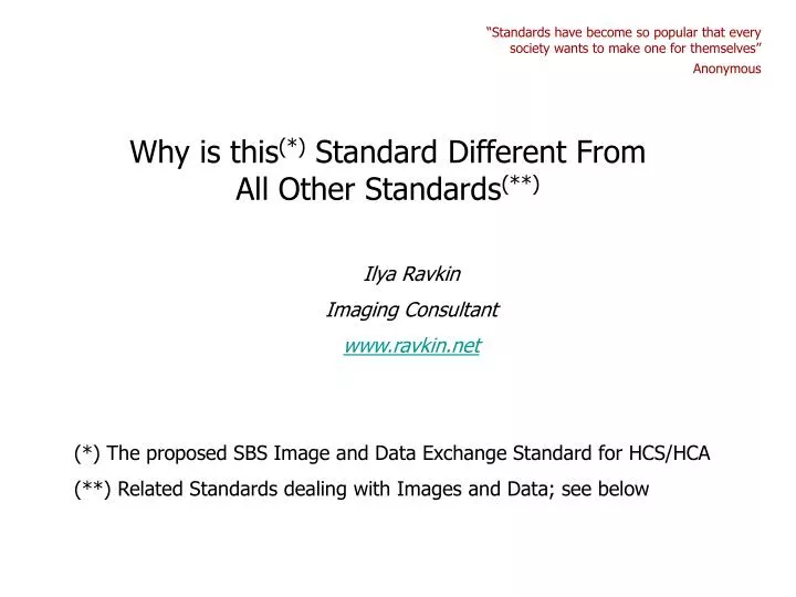
Why is this (*) Standard Different From All Other Standards (**). “Standards have become so popular that every society wants to make one for themselves” Anonymous. Ilya Ravkin Imaging Consultant www.ravkin.net. (*) The proposed SBS Image and Data Exchange Standard for HCS/HCA

E N D
Why is this(*) Standard Different From All Other Standards(**) “Standards have become so popular that every society wants to make one for themselves” Anonymous Ilya Ravkin Imaging Consultant www.ravkin.net (*) The proposed SBS Image and Data Exchange Standard for HCS/HCA (**) Related Standards dealing with Images and Data; see below
Related Standards “The nice thing about standards is that there are so many of them from which to choose” - Andrew Tannenbaum (http://www.sysprog.net/quotes.html) OME - Open Microscopy Environment (http://www.openmicroscopy.org/index.html) MIACA - Minimum Information About a Cellular Assay (http://miaca.sourceforge.net/) Libics - Image Cytometry Standard (http://libics.sourceforge.net) CytometryML (http://www.newportinstruments.com/cytometryml/cytometryml.htm) MIAME - Minimum information about a microarray experiment (http://www.mged.org/Workgroups/MIAME/miame.html) MISFISHIE - Minimum Information Specification For In Situ Hybridization and Immunohistochemistry Experiments (http://mged.sourceforge.net/misfishie/) The Tissue Microarray Data Exchange Specification (http://www.biomedcentral.com/1472-6947/3/5) Bioinformatics Standards for Flow Cytometry (http://flowcyt.sourceforge.net/) FCS - Flow Cytometry Standard (http://www.isac-net.org) Association for Pathology Informatics Standards (http://www.pathologyinformatics.org/standards.htm) DICOM - Digital Imaging and COmmunications in Medicine (http://medical.nema.org/dicom/2003.html) LDIP - Laboratory Digital Imaging Project (http://www.ldip.org/)
OME What is the Purpose of These Standards? - 1 Following are the stated goals of these standards quoted from respective web sites or published articles. “The Open Microscopy Environment (OME) Data Model and XML file: open tools for informatics and quantitative analysis in biological imaging” Genome Biology, 2005, 6:R47 Ilya G Goldberg, Chris Allan, Jean-Marie Burel, Doug Creager, Andrea Falconi, Harry Hochheiser, Josiah Johnston, Jeff Mellen, Peter K Sorger and Jason R Swedlow “To make it possible to manipulate and share image data as readily as genomic data, we are building an image-management system geared to the specific needs of quantitative microscopy.”
MIAME MIACA What is the Purpose of These Standards? - 2 http://www.mged.org/Workgroups/MIAME/miame.html “MIAME describes the Minimum Information About a Microarray Experiment that is needed to enable the interpretation of the results of the experiment unambiguously and potentially to reproduce the experiment.” http://miaca.sourceforge.net “We propose the Minimum Information About a Cellular Assay (MIACA), and develop this as an information guideline and a modular Cellular Assay Object Model (CA-OM) that is capable of covering the range of cellular assays possible and which is the basis for efficient data exchange.”
LDIP MISFISHIE What is the Purpose of These Standards? - 3 MISFISHIE - Minimum Information Specification For In Situ Hybridization and Immunohistochemistry Experiments (http://mged.sourceforge.net/misfishie) “This specification details the minimum information that should be provided when publishing, making public, or exchanging results from visual interpretation-based tissue gene expression localization experiments such as in situ hybridization, immunohistochemistry, reporter construct genetic experiments (GFP/green fluorescent protein, beta-galactosidase), etc.. Compliance to this standard is expected to provide researchers at other labs enough information to reproduce the experiment and/or to fully evaluate the data upon which results are based.” http://www.ldip.org “The Laboratory Digital Imaging Project (LDIP) was developed under the direction of the Association for Pathology Informatics (API) to develop and promote the use of pathology images to improve patient care and improve the quality of research in pathology by the development of standards relating to the interchange of various types of images.”
TMA DES CytometryML What is the Purpose of These Standards? - 4 “The tissue microarray data exchange specification: A community-based, open source tool for sharing tissue microarray data” BMC Medical Informatics and Decision Making 2003, 3:5 Jules J Berman, Mary E Edgerton and Bruce A Friedman “Without such a specification, TMA data files can not be shared or merged, and the full scientific value of this new technology is not realized.” http://www.newportinstruments.com/cytometryml/cytometryml.htm “Cytology automation and research will be enhanced by the creation of a common data format. This data format would provide the pathology and research communities with a uniform way for annotating and exchanging images, flow cytometry, and associated data.”
Purpose of the SBS Image And Data Exchange Standard for HCS/HCA All of the related standards attempt to adequately describe the collected data and facilitate data sharing and pooling among different organizations, hence the names “minimal information about …”. In the academic world and for the advancement of science the comprehensive description of data is necessary and should be enforced either by the standard itself or by some acceptance body, e.g. magazine editors. Our goal at SBS should be not data sharing among different organizations, but interoperability of software from different vendors, which will give greater flexibility to the user. The question of how well or badly the experiment is described and which terms are used for this description should be left entirely in the authority of the user (or the user’s company). This change in purpose has a lot of technical consequences. We do not need to agree on numerous fields, tags, vocabularies, and ontologies. All that is needed, is to agree on the syntax and computer representation. It is almost universally accepted today that XML is the best way to describe this kind of data. The task now becomes quite manageable – to agree on how to parse or update the XML file in order to get access to images, associated with them descriptors, and derived from them numerical results.
Structure and Content Images and Data Imposed by the SBS Image and Data Exchange Standard Structure Content Imposed by each Company/Institution (possibly in compliance with one of the existing standards) Images and Data The Difference Other Standards: This Standard:
After First Data Analysis Measure_1 Not satisfied Dose After Second Data Analysis Measure_2 Dose Example of Information Flow in HCS/HCA Image Acquisition and Image and Data Analysis Company B Data Entry Application (If there was a standard) Company A Imager Before Image Acquisition SampleLayout.xml SamplePlate.xml After Image Acquisition Company C Image Analysis SW SamplePlate.xml After First Image Analysis Company D Data Analysis SW Company E Image Analysis SW SamplePlate.xml After Second Image Analysis Company F Data Analysis SW Satisfied
SampleLayout.xml <PlateLayout> <PlateLayoutID>SAMPLE_LAYOUT</PlateLayoutID> <Well> <WellNum>1</WellNum> <DrugID>LY294002</DrugID> <Dose>0.001</Dose> <CellType>MCF7</CellType> </Well> <!--other wells here --> </PlateLayout> One Well
SamplePlate.xmlAfter Image Acquisition <Plate> <PlateID>SAMPLE_PLATE</PlateID> <PlateLayoutID>SAMPLE_LAYOUT</PlateLayoutID> <Well> <WellNum>1</WellNum> <Channel> <ChannelID>GFP</ChannelID> <ChannelImage>C:\SAMPLE_PLATE\W01-Green.TIF</ChannelImage> </Channel> <Channel> <ChannelID>DRAQ5</ChannelID> <ChannelImage>C:\SAMPLE_PLATE\W01-Red.TIF</ChannelImage> </Channel> </Well> <!--other wells here --> </Plate> One Well
SamplePlate.xmlAfter First Image Analysis <Plate> <PlateID>SAMPLE_PLATE</PlateID> <PlateLayoutID>SAMPLE_LAYOUT</PlateLayoutID> <Well> <WellNum>1</WellNum> <Channel> <ChannelID>GFP</ChannelID> <ChannelImage>C:\SAMPLE_PLATE\W01-Green.TIF</ChannelImage> </Channel> <Channel> <ChannelID>DRAQ5</ChannelID> <ChannelImage>C:\SAMPLE_PLATE\W01-Red.TIF</ChannelImage> </Channel> <Measure> <MeasureName>MEASURE_1</MeasureName> <MeasureMethod>FIRST_METHOD</MeasureMethod> <MeasureMethodParameters>120 0.5 30</MeasureMethodParameters> <MeasureValue>0.73</MeasureValue> </Measure> </Well> <!--other wells here --> </Plate> Added
SamplePlate.xmlAfter Second Image Analysis <Plate> <PlateID>SAMPLE_PLATE</PlateID> <PlateLayoutID>SAMPLE_LAYOUT</PlateLayoutID> <Well> <WellNum>1</WellNum> <Channel> <ChannelID>GFP</ChannelID> <ChannelImage>C:\SAMPLE_PLATE\W01-Green.TIF</ChannelImage> </Channel> <Channel> <ChannelID>DRAQ5</ChannelID> <ChannelImage>C:\SAMPLE_PLATE\W01-Red.TIF</ChannelImage> </Channel> <Measure> <MeasureName>MEASURE_1</MeasureName> <MeasureMethod>FIRST_METHOD</MeasureMethod> <MeasureMethodParameters>120 0.5 30</MeasureMethodParameters> <MeasureValue>0.73</MeasureValue> </Measure> <Measure> <MeasureName>MEASURE_2</MeasureName> <MeasureMethod>SECOND_METHOD</MeasureMethod> <MeasureMethodParameters>60 0.75</MeasureMethodParameters> <MeasureValue>1.24</MeasureValue> </Measure> </Well> <!--other wells here --> </Plate> Added
How to Simplify and Speed up the Adoption of the Proposed SBS Image and Data Exchange Standard for HCS/HCA • Form a permanent SBS Committee to work out technical details. • Provide free and open source SW to read, write, and convert to and from other formats the proposed XML files of HCS/HCA data. • Provide validation data sets in this format, e.g. SBS Image Library for algorithm comparison.
How the Standard Can Change the Business Model for Software Development in HCS/HCA • A biological research group post images, description, and design goals for a new assay (e.g., statistical quality, speed of processing, cost, minimal required number of cells) and relevant restrictions (e.g., type of computer/operating system) and business terms (e.g., needed number of licenses) • Interested image analysis developers compete on this problem and return their results (privately or publicly). • The research group compares results, possibly runs more detailed tests and makes a decision. Both groups sign a contract.