Download
1 / 18
200 likes | 551 Views
Developing A Protein Purification Protocol. Billie Parker 6-14-02. Overview. Introduction to Chromatography Extraction of Protein from Cells Introduction to Green Fluorescent Protein Results Conclusions. Introduction to Chromatography. Chromatography is used to purify complex mixtures.

E N D
Developing A Protein Purification Protocol Billie Parker 6-14-02
Overview • Introduction to Chromatography • Extraction of Protein from Cells • Introduction to Green Fluorescent Protein • Results • Conclusions
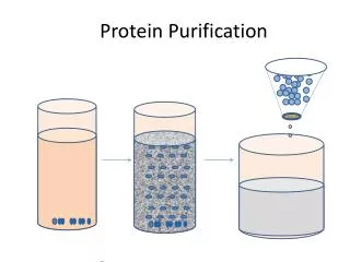
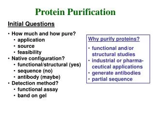
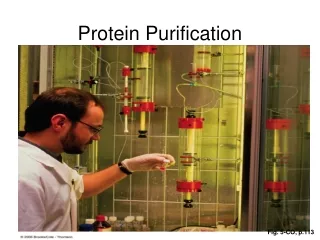
Introduction to Chromatography • Chromatography is used to purify complex mixtures. • We used a resin in a column. • The resin binds to certain proteins. This purifies these proteins away from other proteins that do not bind to the resin. • After the other proteins have passed through, the bound proteins can be released. • There are different resins for different substances you want to purify.
Hydrophobic Interaction • The resin binds to water-hating proteins. • Increased salt concentrations promote binding. • The bound protein can be released by lowering the salt. • We tried three different resins to see which one worked the best.
Column Information • The resin is packed into the column already. • The substance to be purified passes through the resin and binds to it. • Most molecules do not bind to the resin.
FPLC System Components • The buffers carry the sample through the system. • There are two pumps to move the buffers. • Sample is injected into the valve. • Then the sample passes to the column. • The UV light detector measures the amount of protein based on absorbance. • The sample is divided into fractions by the fraction collector.
Extraction of Protein from Cells • Getting protein out of cells is the first step in purification. • We centrifuged the bacteria to get them out of the growth medium. • We used two different methods to break open the bacteria. • We resuspended the cells in detergent. • We resuspended the cells and froze them.
Extraction by Detergent • After lysing the cells in detergent, we centrifuged the cells at 10,000 xg for 10 minutes and kept the supernatant. • We used a particular resin that binds to only detergent to purify the detergent from the supernatant. • We put ammonium phosphate in the extract to increase the amount of salt to promote binding to the resin. • Then, we loaded the extract on the column.
Extraction by Freezing • We used TE Buffer to resuspend the cells. • We put the cultures into the freezer at -70o for 20 minutes. • We let the cells thaw. • Then we centrifuged at 10,000 xg for 10 minutes and kept the supernatant. • We put ammonium phosphate in the extract to increase the amount of salt to promote binding to the resin. • Then, we loaded the extract on the column.
Introduction to GFP • GFP is Green Fluorescent Protein. • GFP is from jellyfish. • We used bacteria that were engineered to make GFP. • GFP glows when you shine UV light on it.
Introduction to GFP • GFP is Green Fluorescent Protein. • GFP is from jellyfish. • We used bacteria engineered to make GFP. • GFP glows when you shine UV light on it.
Results • The graphs will show the amount of protein indicated by UV absorbance. • The salt concentration starts high to allow the GFP to bind then it decreases to let the GFP come out. • GFP was detected by shining UV light on the collected fractions.
First Column Tested • Salt shows the salt concentration in the buffer. • UV measures total protein. • GFP shows fractions that glow.
Second Column Tested • Salt shows the salt concentration in the buffer. • UV measures total protein. • GFP shows fractions that glow.
Third Column Tested • Salt shows the salt concentration in the buffer. • UV measures total protein. • GFP shows fractions that glow.
Conclusions • The octyl column did not bind to the GFP. • Both the phenyl and butyl columns bound the GFP and eluted it with low salt. • We were unable to conclude which column worked the best because we cannot tell how much protein is in the GFP fractions.