Download
1 / 39
600 likes | 1.45k Views
Amino Acid Catabolism II: Fate of Carbon Skeleton. Long-term protein overfeeding accelerates the status insulin resistance. Extra glucose in the fed state. protein rich. Quantitative aspects of amino acids catabolism.

E N D
Amino Acid Catabolism II: Fate of Carbon Skeleton
Long-term protein overfeeding accelerates the status insulin resistance Extra glucose in the fed state protein rich
Quantitative aspects of amino acids catabolism 1. Amino acids only undergo partial oxidation in the liver 2. Partial oxidation ATP in the fed state 3. Hepatic gluconeogenesis 4.Urea synthesis and gluconeogenesis from dietary amino acids on the same pathway. -
Glucogenic and ketogenic amino acids each endproduct can yield a new oxaloacetate can yield FA or ketone body
The 3-Ca-keto acid pyruvate is produced from alanine, cysteine, glycine, serine and partly from tryptophan (indolylalanine). Glucoplastic Alanine via transaminase directly yields pyruvate.
Major pathways of serine in humans glutathion creatine hem bile acids
Serine pyruvate transamination HCO3 3 S-adenosyl-methionine choline Serine ethanolamine H2O neurotransmitter synthesis Serine active C1 transfer Glycine HCO3+NH+4 glycine cleavage transaminase glyoxalate oxalate, transaminase deffect: kidney stones
Sulfur containing amino-acids I. Role in protein structure Methionine – mostly found in the hydrophobic core of proteins, membrane spanning domains, if surface exposed, susceptible to oxidation, e.g.: elastase inhibitor - initiating protein for eukaryotic protein synthesis Cysteine - forms inter-intrachain disulfide bonds with other cysteine residues Cystine
Sulfur-containing amino acid (cysteine, methionine) II. Major pathways Met and Cys incorporate into proteins, homocysteine and taurine do not. Met- essential a.a. 1. 2. re methy lation path way 4 Transmethylation pathway (ubiquitous in all cells) 3. 5. Transsulfuration pathway irreversible (limited expression: liver, kidney intestine,pancreas) MAT: Meth adenosyltransferase SAHH: S-adenosylhomocysteine hydrolase CBS: Cystathionine synthase CGL: Cystathionine lyase MTHFR: Methylenetetrahydrofolate reductase MS: Methionine synthase BHMT: Betaine:homocysteine methyltransferase SHMT: Serine hydroxymethyltransferase 6 antioxidant conjugates with bile
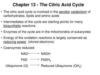
The 4-Coxaloacetate is produced from aspartate and asparagine. Asp - other transamination reactions. Asp - fumarate in the ornithine cycle. Fumarate – oxaloacetate -Asp connects TCA and ornithine cycle. The 4-C TCA cycle intermediate succinyl-CoA is produced from isoleucine, valine, threonin (branched chain amino acids (BCCA) and methionine.
BCCA – branched-chain amino acids: leucine, isoleucine, valine Role in protein structure I. Ile Leu Val Why BCCA? • Nonlinear structure - most hydrophobic amino acids – interior of globular proteins • membranous proteins, surfactants - interaction with phospholipids (lung surfactant protein B) • All essential amino acids (~ 20% BCCA in all dietary proteins) • Stability of folded proteins, effect the folding pathway to form mature protein, thermostability • Function of proteins: create a non-aqueous environment, phospholipid binding, oxygen binding in myoglobin and hemoglobin
BCCA and protein structure II. - Coiled-coiled -helices: fibrinogen, myosine, keratin, transcription factors Leucine-zippers: permit formation of homodimers/heterodimers of transcription factors The bZip family of transcription factors consist of a basic region which interacts with the major groove of a DNA molecule through hydrogen bonding, and a leucine zipper region which is responsible for dimerization.
Tissue distribution of BC aminotransferase and dehydrogenase Ile, Val Gln synthesis BCAA in the diet are metabolized extrahepatically, major site - muscle
Catabolism of BCAA • BCAA metabolism escapes hepatic metabolism • regulatory role in muscle protein • synthesis, insulin secretion, brain amino acid • uptake-leucine. • no unique biologically active degradation • product • catabolized in lockstep • two common steps: BCAT, BCKDH, all 3 • regulated at BCKDH, catabolism is not driven • by the need of glucose or ketone bodies. • - genetic deffect of BCKDH: maple • sirup urine disease (odor of keto acids) Ile Leu Val
Ile, Val Propionyl-CoA - carboxylated to methylmalonyl-CoA. Racemase - L-isomer. Methylmalonyl-CoA Mutase - molecular rearrangement-linear chain of succinyl-CoA. Coenzyme B12 (vitamin B12+ATP, adenosylcobalamine)- cofactor of Methylmalonyl-CoA Mutase.
The 5-C TCA Cycle intermediate a-ketoglutarate is produced from arginine, glutamate, glutamine, histidine and prolin. transport of amino acid N Gla (g- carboxy glutaminic acid)vitamin K GABA (g-amino butyrate) histamin glutamate g-semialdehyde NO creatine glutamate glutamate
histidine lyase Histidine N-formiminoglutamate is converted to glutamate by transfer of the formimino group to THF - N5-formimino-THF. urokanase
C1 unit transfer in amino acid catabolism What is folate? THF+ derivatives
H H 8 7 6 9 5 H H 10 • Tetrahydrofolate (THF), a reduced form of folate. - C1 unit transfer “active carbon” attached to N5 or N10. • C1 units: methyl, methylene, formyl,formimino, methenyl can transform in each other as donors, acceptors. - C1 donated for synthesis of nucleotides, in amino acid metabolism
Interconversion of derivatized THF, role in amino acid metabolism C1 attached to THF
S-Adenosyl-Methionine as methyl donor and its metabolic versatility The methyl group’s transfer at N-5 of THF is insufficient, S-Adenosylmethionine is preferred for methyl transfer methionine adenosyl transferase biotine lipoic acid S+ cal methyl synthesis in plants ,Creatine aminoisopropyl group Liver: SAM is a precursor for glutathione
Bulk of SAM is used in methyltransferase reactions I : • creatine synthesis • phosphatidylcholine synthesis • - 0.6-1.6% of all genes code for methyltransferases at present 25% identified • …….
Transfer of one carbon atom units (C1) histidine glycine serine tryptophan tetrahyrofolic acid (THF4) purine ring thymidilate synthase dTMP formation S-adenosyl S-adenosyl homocysteine methionine “SAM” „CH3-” C1 C1 C1 C1 C1 C1 Vitamin C1 B12 Methyl cycle
Activated methyl cycle (Met and SAM metabolism) Methyltransferase reactions II • SAM: methyl group donor in synthetic reactions, methylation of • DNA, RNA, proteins, biosynthesis of phosphatidylcholine • creatine. • Methyl-H4folate-H4folate S-adenosyl-homocysteine methyltransferase adenosyl transferase diet Homocysteine-methionine Methionine synthase (MS) hydrolase
Methionine metabolism: remarkable vitamin dependence, folate, vitamin B12,B6, FAD Vitamin FAD Vitamin B6 Conversion of homocysteine to methionine is essential to: conserve methionine detoxify homocysteine produce SAM
Regulation of homocysteine formation by SAM level, SAM „switch” Protein (choline, methionine, betaine)- “labil” methyl groups provide methyl group to SAM) Diet: 1-1.5g protein/kg 43% of homocysteine Remethylated 57% transsulfuration • High SAM • Enhance the flow of homocysteine • out of the methionine cycle • Low SAM • Conserve homocysteine within the • methionine cycle
Homocysteine causing oxidative stress Glutathione peroxidase
Homocysteinepotentiates oxidative injury in vascular diseases: coronary artery disease, cerebrovascular events, and in brain degenerative deseases (AD, Parkinson). Genetic predisposition to hyoerhomocysteinemia: most common inherited form of hyperhomocysteinemia: alteration in the gene encoding the enzyme methylene tetrahydrofolate reductase (MTHFR), leading to moderate hyperhomocysteinemia. less often the cause is cystathionine -synthase (CBS) deficiency, with very highhomocysteine levels. Aquired mild hyperhomocysteinemia Dietarydefficiencies of folate, vitamin B12 and/orB6. Cofactors for the optimal function of MTHF and CBS. Vascular diseases Folate recommandation: 400 to 600 μg per day. Insufficient folate – neural tube deffect, spina bifida, anencephaly. Plants vary in their folate level, wheat and rice contain extremely low level - biofortification?
Increased utilisation of SAM due to oxidative stress also results in accumulation of homocysteine • „methyl balance” maintain adequate level of SAM unstable intermediate , aging
Methionine metabolism in the cellular assimilation of folate, the “folate trap” Functions of methionine synthase: 1. methionine conservation 2. cellular folate assimilation by conversation of 5-methyl-THF to THF, to support DNA synthesis. Impaired MS activity (brought about by B12 defficiency): functional folate defficiency. Vitamin B12 is the only acceptor of methyl-THF. There is also only one acceptor for methyl-B12 - homocysteine in a reaction catalyzed by methionine synthase. A defect in homocysteine methyltransferase or a deficiency of B12 can lead to a methyl-trap of THF and a subsequent deficiency. Inhibition of nucleotide synthesis – effecting erythropoiesis – megaloblastic anaemia. (Immature large cells released from the bone marrrow to try to compensate for anemia.) Folate-Diet
Gluco - ketoplastic dioxygenase Tryptophan N-formyl-kynurenin hydroxylase few% formyl (THB) kynurenin formamidase Serotonin (5-hydroxy-tryptamine) THF Regulation of sleeping, psychic processes mood. Serotonin effects accelerated by MAO inhibitors kynurenin Kynureninate accumulation in schizophrenia kynureninase B6 alanine 3-hydroxyantranylate acetoacetyl-CoA nicotinate, NAD+
Tryptophan, and 5-HT (serotonine) and central phatigue 5-HT - arousal, lethargy, sleepiness, mood 5-HT induced central fatigue peripheral exercise
Metabolism of Phe and Tyr Gluco-and ketoplastic Mixed function oxidation one O atom of O2 is reduced to H2O the other is incorporated into amino acid. Tyrosine: precursor of dopamine, epinephrine, norepinephrine.
Genetic deficiency of Phenylalanine Hydroxylase,or defective production of cofactor(cofactor PKU) tetrahydrobiopterine (THB)phenylketonuria(PKU) THB Tyr hydroxilase Phenylpyruvate, phenylacetate and phenyllactate accumulate in blood, urine, damage myelin of nerve cells. 1:10 000 live birth. Mental retardation Treatment: limiting phenylalanine (essential aa) intake. No sweetener - aspartame! (aspartate+phenylalanine) Tyrosine, an essential nutrient for individuals with phenylketonuria, must be supplied in the diet. DOPA dopamine transaminase,dyoxigenase
THB Tyrosine hydroxylase dopamine THB DOPA tyrosinase melanin Tyrosine transaminase Tyrosine: precursor for synthesis of melanins and of cathecolamins. THB is also a cofactor of tyrosine hydroxilase, treatment of cofactor PKU is complicated. High phenylalanine inhibits tyrosinase, on the pathway for synthesis of the pigment melanin from tyrosine. Albinism: deffect of tyrosinase gene
Leucine increasing mTOR signaling in the hypothalamusand regulating food intake L-leucin regulation in the cells of arcuate nucleus (ARC), hypothalamus Amino-acid leucine Leptin Refeeding Glucose, FFA L-leucine (glutamate synthesis) mTOR ~AMPK cholecistokinine Feeding off signal Food intake Leucine: - not synthetized, not metabolized in the liver - its level reflects ingested protein - transported rapidly to neuron and to glia with L- transporters - can selectively stimulate mTOR in the hypothalamus CNS control of energy balance and glucose homeostasis