Download
1 / 26
290 likes | 913 Views
Third Year Organic Chemistry. CO-303 Natural Product Chemistry. Amino Acids, Peptides and Proteins . Convener : Dr. Fawaz Aldabbagh. Primary, Secondary, Tertiary and Quaternary structures of Proteins.

E N D
Third Year Organic Chemistry CO-303 Natural Product Chemistry Amino Acids, Peptides and Proteins Convener : Dr. Fawaz Aldabbagh Primary, Secondary, Tertiary and Quaternary structures of Proteins. Isoelectric Point. Prosthetic Group. Investigation of amino acid structure of a protein. Peptide Synthesis
Amino Acids All DNA encoded aa are All are chiral, except Glycine R = H All DNA encoded aa are usually L-
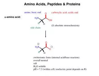
aa are high melting point solids! Why? Answer = aa are ionic compounds under normal conditions Isoelectric Point = concentration of zwitterion is at a maximum and the concentration of cations and anions is equal For aa with basic R-groups, we require higher pHs, and for aa with acidic R-groups, we require lower pHs to reach the Isoelectric Point
Isoelectric Pointis the pH at which an aa or peptide carries no net charge. i.e. [RCOO-] = [RNH3+] So, for basic R-groups, we require higher pHs, and for acidic R-groups we require lower pHs e.g. Isoelectric point for gly pH = 6.0 Asp pH = 3.0 Lys pH = 9.8 Arg pH = 10.8
Preparation of Amino Acids The Strecker Reaction Preparation of Optically active Amino Acids - (Asymmetric Synthesis)
Resolution Prepare the target aa in racemic form, and separate the enantiomers afterwards 1. Crystallisation with a chiral Counter-ion Strechnine
2. Form Diastereotopic Peptides 3. Chiral HPLC 4. Enzyme Resolution Form the N-ethanoyl (acetyl) protected aa then treat with an acylase enzyme.
aa are covalently linked by amide bonds (Peptide Bonds) The resulting molecules are called Peptides & Proteins • Features of a Peptide Bond; • Usually inert • Planar to allow delocalisation • Restricted Rotation about the amide bond • Rotation of Groups (R and R’) attached to the amide bond is relatively free
aa that are part of a peptide or protein are referred to as residues. Peptides are made up of about 50 residues, and do not possess a well-defined 3D-structure Proteins are larger molecules that usually contain at least 50 residues, and sometimes 1000. The most important feature of proteins is that they possess well-defined 3D-structure. Primary Structureis the order (or sequence) of amino acid residues Peptides are always written and named with the amino terminus on the left and the carboxy terminus on the right
Cysteine residues create Disulfide Bridges between chains This does not reveal Primary Structure
Prof. Linus Pauling Dr. Frederick Sanger, Nobel Prize for Chemistry 1958 and 1980 Peptide sequencing Prof. R. B. Merrifield Nobel Prize for Chemistry 1984 Automated Peptide Synthesis
Secondary Structure The Development of Regular patterns of Hydrogen Bonding, which result in distinct folding patterns -helix -pleated sheets
Tertiary Structure This is the 3D structure resulting from further regular folding of the polypeptide chains using H-bonding, Van der Waals, disulfide bonds and electrostatic forces – Often detected by X-ray crystallographic methods Globular Proteins– “Spherical Shape” , include Insulin, Hemoglobin, Enzymes, Antibodies ---polar hydrophilic groups are aimed outwards towards water, whereas non-polar “greasy” hydrophobic hydrocarbon portions cluster inside the molecule, so protecting them from the hostile aqueous environment ----- Soluble Proteins Fibrous Proteins – “Long thin fibres” , include Hair, wool, skin, nails – less folded ----- e.g. keratin - the -helix strands are wound into a “superhelix”. The superhelix makes one complete turn for each 35 turns of the -helix.
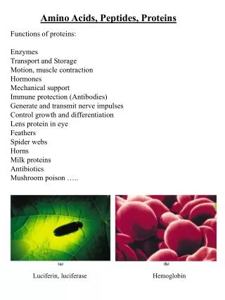
In globular proteins this tertiary structure or macromolecular shape determines biological properties Bays or pockets in proteins are called Active Sites Enzymes are Stereospecific and possess Geometric Specificity The range of compounds that an enzyme excepts varies from a particular functional group to a specific compound Emil Fischer formulated the lock-and-key mechanism for enzymes All reactions which occur in living cells are mediated by enzymes and are catalysed by 106-108 Some enzymes may require the presence of a Cofactor. This may be a metal atom, which is essential for its redox activity. Others may require the presence of an organic molecule, such as NAD+, called a Coenzyme. If the Cofactor is permanently bound to the enzyme, it is called a Prosthetic Group.
For a protein composed of a single polypeptide molecule, tertiary structure is the highest level of structure that is attained Myoglobin and hemoglobin were the first proteins to be successfully subjected to completely successful X-rays analysis by J. C. Kendrew and Max Perutz (Nobel Prize for Chemistry 1962) Quaternary Structure When multiple sub-units are held together in aggregates by Van der Waals and electrostatic forces (not covalent bonds) Hemoglobin is tetrameric myglobin For example, Hemoglobin has four heme units, the protein globin surrounds the heme – Takes the shape of a giant tetrahedron – Two identical and globins. The and chains are very similar but distinguishable in both primary structure and folding
Protecting Groups Protecting NH2