Download
1 / 59
970 likes | 1.87k Views
Why Suffer from. Heel Pain. Brian K. Wagner, DPM, CWS St. Francis Hospital and Medical Center. Education. 4 Years of Undergraduate Studies 4 Years of Podiatry School 1-3 Years of Residency Training concentrating on podiatric medicine and surgery. Brian K. Wagner, DPM, CWS.

E N D
Why Suffer from Heel Pain Brian K. Wagner, DPM, CWS St. Francis Hospital and Medical Center
Education • 4 Years of Undergraduate Studies • 4 Years of Podiatry School • 1-3 Years of Residency Training concentrating on podiatric medicine and surgery
Brian K. Wagner, DPM, CWS • Graduated high school in 1996 in New York. • Three years at C.C.S.U. on advanced academic scholarship. • Podiatry school at Chicago Medical School/ Scholl College of Podiatric Medical School. • Completed three years of residency at St. Francis Hospital and Medical Center. • Two of the three years as Chief Resident. • Certified Wound Care Specialist.
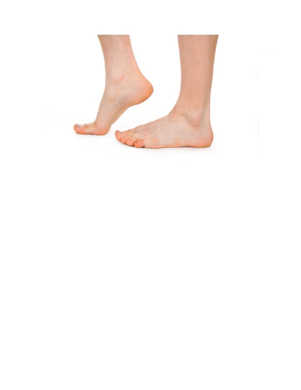
Anatomy • Plantar Fascia • A fibrous band • Supports the arch
Anatomy of the Plantar Fascia • Broad, dense band of longitudinally arranged collagen fibers • 3 bands: medial, central, lateral • Origin: anterior aspect of calcaneal tuberosity • Distally divides into 5 digital bands at the metatarsophalangeal joints • Each digital band pass on either side of flexor tendons and inserts dorsally at the base of the toes.
Anatomy • Posterior Tibial Nerve • Supplies bottom of foot and heel. • Flexor Retinaculum • Supports Tendons, Arteries, and Nerves • Nerve compression may occur.
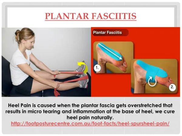
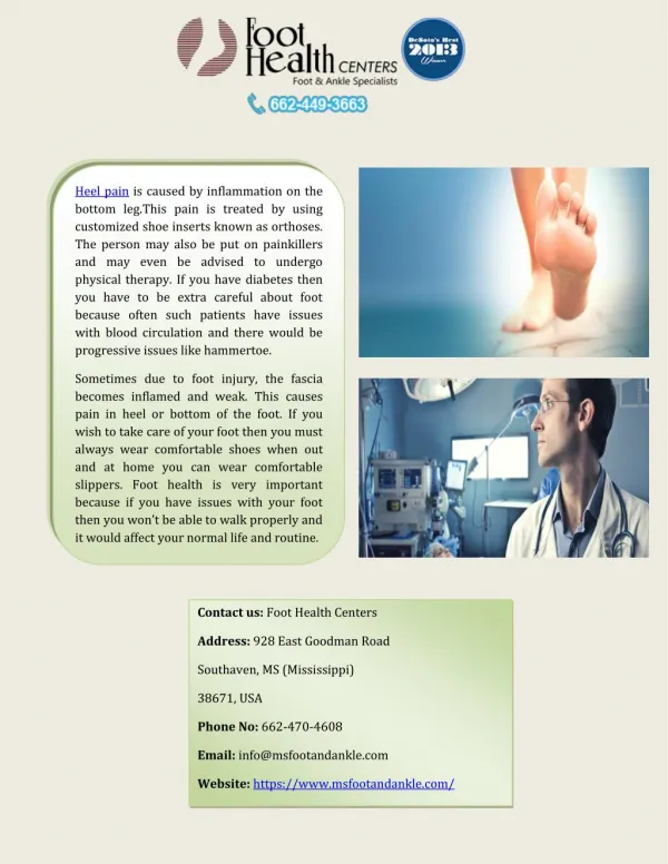
Plantar Fascia • Ligament that supports the foot and arch. • Helps in shock absorption. • As the foot flattens there is a stretch on the ligament causing inflammation and tearing.
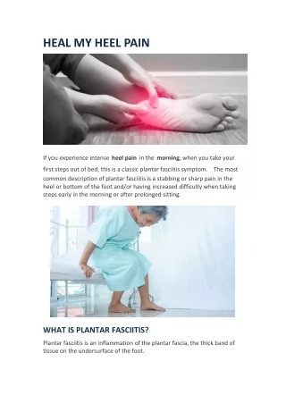
Symptoms • First step pain. • Feels like a pulling sensation when walking. • Pain possibly worse while barefoot or wearing a low-heeled or unsupportive shoe.
Physical Examination • Majority-pain upon palpation of the medial tubercle of the calcaneus* • Palpatory tenderness extends into the medial arch • Pain may be elicited by extension of foot
Heel Spur • Chronic strain on plantar fascia at attachment to heel bone, creates small tears -> causing new bone formation=“heel spur”. • Heel spur=“fishhook-shaped structure” • Pain often due to inflammation not spur.
Diagnosis • History • Pain first step in the morning. • Pain after sitting. • Pain on the bottom of the heel. • Morning limping. • Physical • Pain localized to the plantar fascia insertion. • Pain along plantar fascia with toes flexed.
plantar fasciitis inflammation of tendon or bursa heel bruise systemic disease such as rheumatoid arthritis nerve entrapment heel bone stress fracture tumor plantar fascia rupture soft tissue infection bone infection neuritis fat pad syndrome infracalcaneal bursitis Differential Diagnoses
X-rays • May or may not show anything • May show a heel spur. • Can help rule out other diagnoses.
Further Studies • MRI • Ultrasound • Bone Scan • EMG • Laboratory Studies (ESR, serum urate, rheumatoid panel, etc.)
Biomechanical Causes repetitive pull or strain on soft tissue attachments foot type (low or high arch) obesity inappropriate shoe gear unyielding surfaces extensive activity varus/valgus foot type Systemic Causes Rheumatoid Arthritis Gout Reiter’s Syndrome Ankylosing Spondylitis DISH Infection Causes of Heel Pain:
Most common biomechanical cause of heel pain • tight heel cord • fibrous band relaxes and tightens • tightness of Achilles tendon and fibrous band produces strain and possibly injury • may result in tears within fascia and even possibly bone spur • depending on foot type, plantar fascia is tight already or becomes tight
High Arch Foot Type • Pes Cavus=high arch • not good at shock absorption • already tight plantar fascia • produces additional strain from forces while walking
Low Arch Foot Type • Pes Planus=low arch foot • plantar fascia BECOMES tight • unstable foot • arch collapses with each step • walking lengthens foot and adds plantar-fascial strain
Heel Pain Treatment • 80-95% of patients treated successfully with conservative treatment • 2 Goals: • Calm down current inflammation • prevent inflammation from coming back
Conservative Treatment Padding Proper shoes Reduce activity Muscle stretching Orthotics RICE principles NSAIDs Steroid Injection ESWT – sound waves Physical Therapy Night splints Conservative care is successful 85% of the time. Plantar Fasciitis
Plantar Fasciitis • Surgical Treatment • Ligament Release • Ligament Resection • Recommend orthoses after surgery to prevent recurrence.
Arch taping or padding • for acute onset pain • can be used as only treatment • possibly used as trial regarding orthotic benefits • Advantages: minimal cost, quick and easy to apply
What you can do at home • Change Shoe gear • good arch and support • Ice Pack or Massage • 20 minutes on-off • Rest • Over-the-counter anti-inflammatory medicine (if tolerated) • Stretching and strengthening exercises
What your doctor can do • Prescribe orthotics • over the counter • acute pain • low cost • custom molded orthotics • controls biomechanical risk factors • up to 80% effectiveness • low arch • high arch • excessive motion
What your doctor can do • Prescribe Physical Therapy and/or Home Exercise Program • Stretch Achilles tendon and plantar ligament • ex: wall stretches, rolling foot over tennis ball, towel stretching • Strengthen weak intrinsic foot muscles • ex: towel curls, coin/marble grasping, toe taps
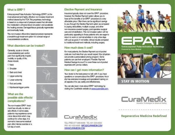
Extracorporal Shock Therapy • Indicated for heel pain over 6 months unresolved with conservative therapy • Not indicated for arch pain. Pain located at the tubercles • Generate high energy pulses 2-3 times/second • Causes “neovascularization”- new blood vessels promote healing
Extracorporal Shock Therapy • 2 devices with FDA approval: • Ossatron Orthotripsy • Dornier Epos
Other available options • Posterior night splints • passively stretches calf, plantar fascia, and Achilles tendon while sleeping • decreases AM tension • Disadvantages: • possible mild discomfort • possible sleep interruption
Surgical Treatments • reserved only for patients not responding to conservative therapy • involves cutting of plantar fascia and possible removal of heel bone spur
Endoscopic Plantar Fasciotomy • closed approach through small incision • cutting medial 1/3 to 1/2 of fibrous band • advantages: less trauma, less postop pain, quicker recovery, and can bear weight